Targeting antigens to dendritic cells (DCs) seems like a logical strategy for cancer immunotherapy, but without co-administration of an adjuvant to activate target DCs, this strategy has shown little efficacy. In a recent paper, published in the Journal of Clinical Investigation, Zeng et al. describe the self-adjuvanting effect of a nanoemulsion delivery system that targets antigen to Clec9A, a key endocytic receptor on CD8+ cross-presenting DCs, to stimulate a potent, antigen-specific antitumor immune response.
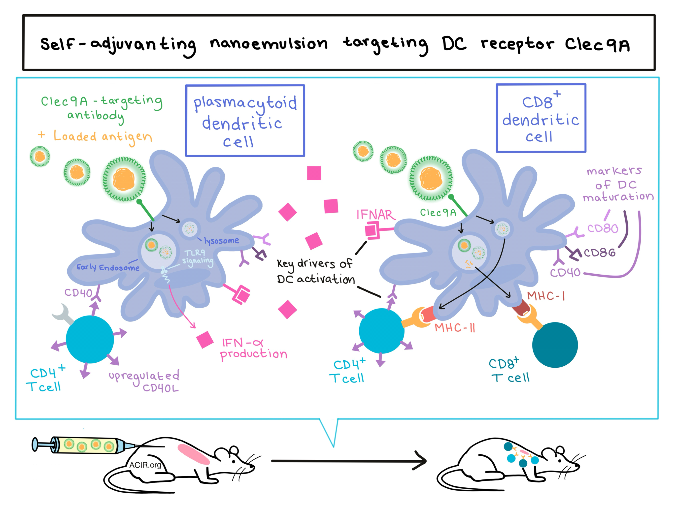
In a previous study, Zeng et al. developed functionalized, Clec9A-targeting tailored nanoemulsions (Clec9A-TNE) using an antibody specific for murine Clec9A. The oil-in-water nanoemulsion system incorporates and delivers antigens directly to Clec9A, which is responsible for organizing the processing and cross-presentation of antigens as epitopes on MHC-I. Clec9A is present on cross-presenting CD8+ DCs and on plasmacytoid DCs (pDCs) in mice. When OVA was delivered via the nanoemulsion, it induced antigen-specific CD4+ and CD8+ T cell responses in vitro, encouraging the current in vivo study and mechanistic investigation of the immunogenicity of this approach.
To better understand the behavior of their nanoparticles, the researchers utilized fluorescently labeled Clec9A-targeted and untargeted TNE. In vivo, they observed early accumulation in both the spleen and liver, but by day 7, the Clec9A-targeted TNE remained in the spleen only, while non-targeted TNE were found in the liver only. In vitro, they examined intracellular localization and found that after recognition by the Clec9A receptor, antigen-loaded TNE are internalized by dendritic cells and reach both lysosomes and early endosomes. Presence of antigen in the early endosome compartment is considered critical for DC cross-presentation.
The researchers next tested the ability of OVA-Clec9A-TNE to induce an immune response in vivo. In addition to induction of a strong anti-OVA antibody response, they found that the OVA-Clec9A-TNE induced strong proliferation of adoptively-transferred, OVA-specific CD4+ and CD8+ T cells, and that these activated T cells were capable of killing OVA-expressing target cells. Given the importance of DC activation in generating an immune response, the researchers explored the activation status of DCs after OVA-Clec9A-TNE administration and observed that all splenic DC subsets (CD8+ DCs, CD8- DCs, and pDCs) showed increased expression of DC maturation markers CD86, CD80, and CD40 following administration of OVA-Clec9A-TNE. Since Clec9A only targets CD8+ and pDCs, the upregulation of maturation markers on CD8- DCs indicated that something other than direct targeting by the nanoemulsion had led to the broad DC activation.
Two key mechanisms of DC activation engendered by the nanoemulsion emerged which were critical to effective CD8+ T cell stimulation. First, OVA-Clec9A-TNE induced systemic production of IFN-α, while all other tested constructs, including Clec9A-targeted OVA protein together with the adjuvant CpG, did not. Later data showed that the IFN-α was released by pDCs in a TLR9- and CD40-dependent manner and that TLR9, the IFN-receptor, and the systemic production of IFN-α are critical to the nanoemulsion system’s ability to act as a self-adjuvant. Second, the requirement for CD4+ epitopes in the Clec9A-targeted TNE, the upregulation of CD40L on CD4+ T cells, and the absence of an effect in CD40-/- mice indicate that CD4+ T cell help was also crucial to full activation of DCs by the Clec9A-targeted nanoemulsion.
To test whether the immunogenicity of their system would translate to antitumor efficacy, the researchers tested the OVA-Clec9A-TNE in mice with OVA-expressing breast cancer tumors; treated mice showed significantly inhibited tumor growth and prolonged survival compared to controls. Antitumor efficacy was found to correlate with greater infiltration by inflammatory DCs, CD4+ T cells, and CD8+ T cells.
To address the issue that an antibody-mediated targeting system might not hold up in long-term use in vivo, which would be required against more realistic antigen targets, the team showed that two other Clec9A-targeting approaches – a peptide selected by phage display to bind Clec9A (WH) and the natural Clec9A ligand polymerized actin (F-actin) – were equivalent to the antibody to Clec9A in forming immunogenic emulsions. Using WH-TNE, the team tested the delivery of the HPV-associated E6/E7 antigen and found that it effectively suppressed growth of HPV-associated TC1 tumors, induced strong E6/E7-specific IFNγ responses, and prolonged survival.
To test whether they could target neoantigens, the researchers next loaded nanoemulsions with 18 previously described neoepitopes for the B16-F10 melanoma model. They conducted an initial test to evaluate each neoepitope for immunogenicity based on the reaction elicited from splenocytes, and identified a preferred stimulatory pool of 6 neoepitopes. When WH-TNE encapsulating this stimulatory neoepitope pool was delivered to B16-F10 melanoma-bearing mice, it induced a neoepitope-specific IFNγ response, significant suppression of tumor growth, and enhanced survival. The antitumor effects were CD4+ T cell dependent, and all tumor-free mice previously treated with WH-neoepitope-TNE were protected against a B16-F10 melanoma rechallenge, indicating a protective memory response.
Overall, the results of this study show that this Clec9A-targeting nanoemulsion delivery system can target antigen to CD8+ cross-presenting DCs, ultimately stimulating a potent, antigen-specific antitumor response by CTLs. In the future, this strategy could be tailored to individual patients and applied as a personalized cancer immunotherapy.
by Lauren Hitchings


