Weekly Digests
‹ Back to March

TAMs may be friends, not foes, in early-stage tumor T cell response

March 6, 2019

Knowing that immunotherapy that works in mice often fails in patients with solid tumors, and that mouse models mostly utilize tumor cell lines from advanced tumors, Singhal et al. decided to bypass murine studies entirely and instead analyzed the functions of macrophages and monocytes and their interactions with tumor-specific T cells within early-stage human lung tumors. The results, recently published in Science Translational Medicine, demonstrated that the microenvironment in early-stage cancers is quite different from the microenvironment in advanced tumors, suggesting that different immunotherapy strategies may be necessary for the treatment of various tumor stages.

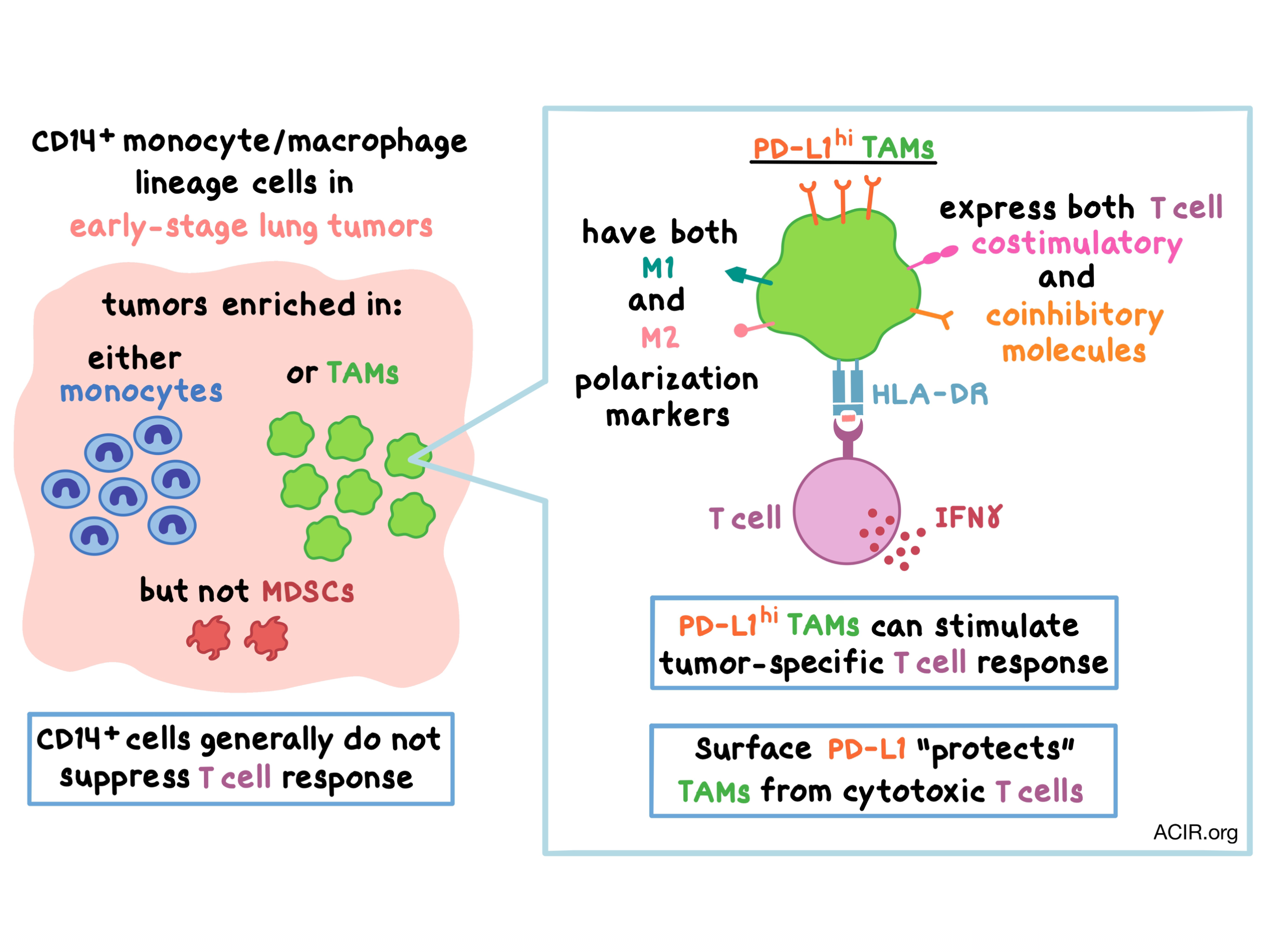
To begin, the researchers phenotypically profiled immune cells from lung cancer tissue, adjacent lung parenchyma, and peripheral blood in 93 patients with stage 1 or 2 non-small cell lung cancer (NSCLC). They found that most CD45+CD11b+ myeloid cells within the tumor were either CD14+ monocyte/macrophage lineage cells (MMLCs) or CD66b+ granulocytic lineage cells. The team observed that the MMLCs did not preferentially accumulate in the tumors. Based on markers and morphology, Singhal et al. determined that CD14+HLA-DRhi cells were tumor-associated macrophages (TAMs) and CD14+HLA-DRint cells were monocytes. Many tumors were enriched in TAMs or monocytes, however, there was only a low frequency of CD14+HLA-DRlo/- monocytic myeloid-derived suppressor cells (MDSCs) present in the early-stage tumors.

Next, Singhal et al. observed that the CD14+ cells within the tumor, and to a lesser extent, the adjacent lung tissue, had high expression of both T cell co-inhibitory and costimulatory molecules. Moreover, TAMs co-expressed both M1 (pro-inflammatory) and M2 (anti-inflammatory, pro-tumor) polarization markers in the early-stage tumors. Surprisingly, PD-L1 expression in TAMs was correlated with expression of T cell costimulatory molecules, and the tumors were enriched in PD-L1hi TAMs.

The team then analyzed the interactions between tumor-associated MMLCs and T cells. In an antigen-nonspecific assay, tumor-derived CD14+ cells generally did not suppress autologous T cell proliferation or IFNγ production, and in some cases, they even exerted a stimulatory effect on the T cells. To study the effect of MMLCs on tumor-specific T cell responses, the researchers utilized an in vitro model system comprising human T cells transduced with Ly95 – a high-affinity transgenic TCR recognizing an HLA-A*0201-restricted NY-ESO-1 peptide – and A549 human lung adenocarcinoma cells modified to express the NY-ESO-1 peptide. Coculture of these two cell types led to Ly95 T cell activation and production of IFNγ and granzyme B. The addition of tumor-derived CD14+ cells to the coculture had minimal effect on Ly95 T cell response for most patients, although in some cases, either suppressive or stimulatory effects were observed. These effects were mostly antigen-specific and required direct cell-to-cell contact. The researchers sought to elucidate the source of the discrepancies between patients, but found no correlation between CD14+ cell-mediated suppressive effects and M2 macrophage marker expression or MDSC presence.

The researchers then examined the effect of tumor PD-L1 expression on T cell function by co-culturing Ly95 T cells with the target tumor cells that were modified to express PD-L1. Not surprisingly, tumor cell PD-L1 expression inhibited T cell activation, cytolytic activity, and IFNγ production, and these effects were mostly reversed by anti-PD-1. In contrast, PD-L1+CD14+ cells (with or without anti-PD-L1) generally did not affect T cell responses, although PD-L1hi TAMs sometimes stimulated the T cells. To study this further, the team loaded TAMs with long peptides specific for the Ly95 T cells and confirmed that these TAMs directly stimulated cognate T cell responses despite high PD-L1 expression. Further experiments demonstrated that in the event of cognate tumor antigen cross-presentation, high PD-L1 expression on TAMs protected them from tumor-specific T cell killing. Thus, paradoxically, anti-PD-1 treatment may limit the immune stimulatory effect of TAMs, diminishing the antitumor effect in early-stage tumors.

Overall, Singhal et al. bring to light and begin to unravel the dynamic and complex roles that MMLCs may have in various stages of tumor development. CD14+ MMLCs in early-stage NSCLC tumors generally did not affect T cell response in vitro, and the accumulation of MMLCs, TAMs, and monocytes in the tumors did not significantly affect the overall survival of the patients. The absence or presence of PD-L1 on CD14+ MMLCs did not alter their effect on T cell response; however, PD-L1hi TAMs sometimes stimulated tumor-specific T cells, whereas PD-L1+ tumor cells suppressed T cell effector function. The immune stimulatory effect of PD-L1hi TAMs, coupled with the co-expression of M1 and M2 polarization markers on TAMs in early-stage lung tumors, underscores the importance of understanding the tumor microenvironment at different stages and has implications for immunotherapy design.

by Anna Scherer

References:

Singhal S., Stadanlick J., Annunziata M.J., Rao A.S., Bhojnagarwala P.S., O'Brien S., Moon E.K., Cantu E., Danet-Desnoyers G., Ra H.J., Litzky L., Akimova T., Beier U.H., Hancock W.W., Albelda S.M., Eruslanov E.B. Human tumor-associated monocytes/macrophages and their regulation of T cell responses in early-stage lung cancer. Sci Transl Med. 2019 Feb 13.

In the Spotlight...

FLEXamers: A Double Tag for Universal Generation of Versatile Peptide-MHC Multimers

With the goal of being able to perform antigen-specific T cell identification, T cell isolation, and TCR avidity characterization using only one type of pMHC precursor for all uses, Effenberger, Stengl, and Schober et al. designed “FLEXamers” – novel, double-tagged pMHC constructs that allow for reversible multimerization via a Strep-tag and versatile functionalization with any probe (biotin, fluorophore, DNA-oligonucleotides, etc.) via a Tub-tag. FLEXamers performed equivalently with currently used methods, but were generated via a much simpler process, suggesting potential utility in personalized immunotherapy approaches.

Atypical motifs in the cytoplasmic region of the inhibitory immune co-receptor LAG-3 inhibit T cell activation

Using T cells that expressed full-length or mutant (deletion or single alanine substitution) LAG-3, and an anti-LAG-3 antibody that efficiently prevents LAG-3 from binding to its ligand, peptide-loaded MHCII, Maeda and Sugiura et al. found that LAG-3-mediated inhibition of T cell activation (IL-2 production) correlated with LAG-3 cell surface expression levels and that LAG-3 transduces two independent intracellular inhibitory signals: through a motif in the membrane-proximal region, deemed Fxxl, and through a C-terminal EX repeat. These newly identified motifs suggest a distinct inhibitory mechanism for LAG-3 compared with other known inhibitory receptors.

IDO1 inhibition potentiates vaccine-induced immunity against pancreatic adenocarcinoma

In a pancreatic adenocarcinoma mouse model, a GM-CSF-secreting pancreatic tumor cell vaccine (GVAX) induced CD4+ and CD8+ T cell infiltration, but also induced IDO1 expression on tumor epithelial cells and in lymphoid aggregates, correlating with worse survival. The addition of an IDO1 inhibitor enhanced effector T cell function and survival. Mechanistically, the IDO1 inhibitor led to suppression of Tregs, reduced capacity of MDSCs to suppress T cell proliferation, and Th1 and Th17 polarization of CD4+ T cells. The addition of anti-PD-L1 had no additional effect and may have detracted from the effect of IDO1 inhibitor and GVAX.

Neoadjuvant nivolumab modifies the tumor immune microenvironment in resectable glioblastoma

Schalper, Rodriguez-Ruiz, and Diez-Valle et al. describe a phase II clinical trial of 29 first-line or salvage resection glioblastoma patients receiving single-dose neoadjuvant nivolumab followed by resection and adjuvant nivolumab. Compared to an historical control cohort not receiving immunotherapy, neoadjuvant nivolumab increased chemokine mRNA expression and prevented reduction of immune markers in IHC. Infiltrating CD8+ T cells expressed high CD69 but low 4-1BB by flow cytometry, and TCR sequencing found a more diverse TCR repertoire correlating with increased survival. All 3 primary surgery cases showed remarkably long survival (ongoing).

Contributed by Alex Najibi

A T-cell-engaging B7-H4/CD3 bispecific Fab-scFv antibody targets human breast cancer

Iizuka et al. designed and manufactured bispecific antibodies (BsAbs) targeting CD3 and B7-H4 (an immune checkpoint molecule overexpressed in multiple cancers) in a Fab-scFv format. Bypassing peptide-MHC TCR recognition, anti-B7-H4/CD3 BsAbs led to lymphocyte-mediated lysis of B7-H4+ breast cancer cells in vitro. In a humanized mouse model, anti-B7-H4/CD3 BsAbs increased tumor infiltration by cytotoxic lymphocytes and reduced tumor volume; this was mediated mostly by CD8+ T cells and partially by CD4+ T cells. High B7-H4 expression was frequently observed in human breast and ovarian cancer, independent of HER2 or PD-L1.

Phase Ib evaluation of a self-adjuvanted protamine formulated mRNA-based active cancer immunotherapy, BI1361849 (CV9202), combined with local radiation treatment in patients with stage IV non-small cell lung cancer

In a phase 1b clinical trial, BI1361849 – made up of sequence-optimized mRNA encoding six NSCLC-associated antigens formulated with protamine – was tested in combination with local radiation in 26 stage IV NSCLC patients who had PR or SD following first-line therapy; 73% of patients continued on second-line maintenance treatment. Treatment was well tolerated and 84% of patients showed antigen-specific humoral and/or cellular immune responses against one or more BI1361849 antigens. One patient achieved partial response, 46% of patients achieved stable disease, and shrinking of some non-irradiated lesions was observed.

REVIEW: Macrophages as regulators of tumour immunity and immunotherapy

Denardo and Ruffell review the role of tumor-associated macrophages in tumor immunity. This heterogeneous myeloid population can be exploited by many factors in the TME (hypoxia, fibrosis, tumor- and lymphocyte-derived signals) to promote angiogenesis, immunosuppression, and metastasis. They explore the direct and indirect mechanisms by which macrophages suppress antitumor immune functions or prevent immune cell infiltration, as well as ways to therapeutically target macrophages (depletion, prevention of recruitment, repolarization) to improve antitumor immunity.

T cells genetically engineered to overcome death signaling enhance adoptive cancer immunotherapy

Upon finding that the apoptosis-inducing ligand FasL is overexpressed in most cancer types, and that its receptor Fas is highly expressed on T cells used in clinical adoptive cell transfer, Yamamoto et al. transduced T cells with dominant negative Fas variants that do not recruit FADD (Fas-associated via death domain), disrupting apoptotic signaling. T cells with engineered CAR or TCR and an overexpressed mutant Fas variant were protected from apoptosis in vitro, and showed enhanced persistence and antitumor effect upon adoptive transfer in mice with solid or liquid tumors, without inducing abnormal lymphoproliferation or autoimmunity.

Everything New this Week In...

Close Modal

Small change for you. Big change for us!

This Thanksgiving season, show your support for cancer research by donating your change.

In less than a minute, link your credit card with our partner RoundUp App.

Every purchase you make with that card will be rounded up and the change will be donated to ACIR.

All transactions are securely made through Stripe.