Blockade of the PD-1/PD-L1 axis effectively reverses T cell exhaustion in some instances of cancer, but understanding what defines “exhaustion” and which cells have the potential for reinvigoration will be important in predicting responses, improving efficacy, and broadening the applications of PD-1 checkpoint blockade. To better understand T cell exhaustion in the context of cancer, Thommen et al. evaluated intratumoral CD8+ T cell populations from patients with non-small-cell lung cancer (NSCLC) and categorized tumor-infiltrating lymphocytes (TILs) as having high PD-1 expression (PD-1T), intermediate PD-1 expression resembling that of healthy donor PBMCs (PD-1N), or no PD-1 expression (PD-1-), and compared their transcriptional, metabolic, and functional signatures.
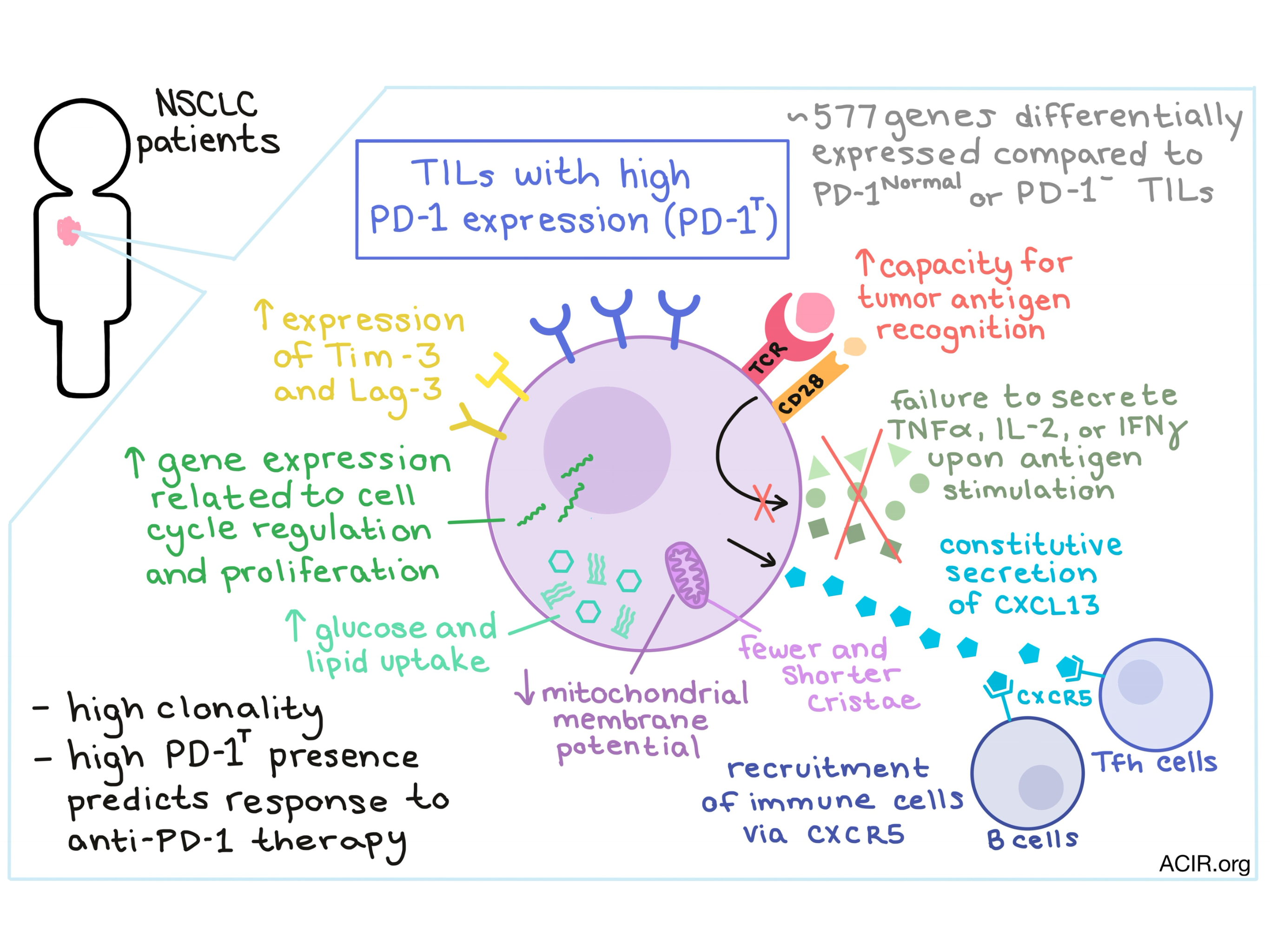
First working to define characteristics of the the PD-1T subset, Thommen et al. noted that PD-1T TILs showed upregulation of additional exhaustion markers, including Tim-3 and Lag-3. Ex vivo assessment of cytokine secretion following CD3/CD28 stimulation showed that PD-1T TILs failed to produce substantial amounts of classical effector cytokines including IL-2, TNFα, or IFNγ. The PD-1T subset was also characterized by high clonality, with the top 30 clones accounting for nearly 90% of the TCR repertoire, suggesting that this phenotype might be driven by TCR signaling. Further, these dominant clonotypes were not frequent among PD-1N or PD-1- cells. Interestingly, following in vitro expansion, PD-1T TILs reduced PD-1 levels, regained effector function, and were found to have an increased capacity for tumor recognition, with tumor reactivity largely restricted to the PD-1T subset.
Using RNA-seq, Thommen et al. uncovered that while PD-1N and PD-1- populations had nearly overlapping gene expression profiles, with only 8 genes differentially expressed, cells within the PD-1T subset had a vastly different transcriptional signature from PD-1N TILs, with 577 genes significantly up- or downregulated between them. Among the genes most upregulated in PD-1T TILs were those related to cell cycle regulation and proliferation (deviating from what has been observed in chronic infection models), and cell cycle analysis showed that PD-1T TILs were able to progress through the cell cycle. While PD-1T TILs were highly clonal and expressed high levels of inhibitory receptors and reduced levels of activating receptors, they showed little evidence of senescence.
Investigating whether high PD-1 expression by TILs could be linked to metabolic changes, the researchers found that cells in the PD-1T subset showed signs of increased glucose and lipid uptake and high lipid content, which could possibly be accounted for by observed expression of the scavenger receptor CD36. PD-1T TILs had, overall, a slightly larger mitochondrial mass, however, the mitochondrial membrane potential was found to be significantly reduced, indicating dysfunctionality. Morphological changes including fewer and shorter cristae were also observed.
When PD-1T TILs were expanded in vitro, derivative TILs continued to express high levels of Tim-3 and Lag-3, which could indicate an underlying, inherited state of dysfunction. Having observed that monocytes and tumor cells in NSCLC both express high levels of IL-10 and that the number of IL-10-producing cells correlated with the number of PD-1T TILs, the researchers tried exposing IL-2-expanded TILs to IL-10 and found that PD-1T-derived TILs rapidly re-expressed high levels of PD-1. Expansion in the presence IL-10 reduced IFNγ production most severely in PD-1T TILs. Together, these results indicate that the PD-1T subset might be inherently susceptible to certain modes of immunosuppression.
Early cytokine secretion analysis had shown that PD-1T TILs failed to produce high levels of classical effector cytokines, but interestingly, transcriptome analysis revealed that they did constitutively secrete high levels of the effector chemokine CXCL13, which controls recruitment and organization of B cells within lymphoid follicles. Through in vitro testing, the researchers found that CXCL13 secretion by PD-1T TILs was able to to attract CXCR5 receptor-expressing cells including B cells, CD4+ follicular helper T cells (Tfh), and a subset of exhausted CD8+ T cells, with the migration of CD4+ T cells being the strongest. In vivo, PD-1T TILs localized in intra- and peritumoral tertiary lymphoid structures alongside Tfh and B cell infiltrates, indicating that PD-1T may play an active role in recruitment of these immune cell types to the tumor.
Finally, Thommen et al. evaluated the presence of PD-1T TILs in pretreatment biopsies from twenty-one NSCLC patients who were being treated with PD-1 blockade therapy and found that the presence of PD-1T TILs strongly predicted response to therapy and correlated with durable responses and longer overall survival.
These studies clarify the qualities of “exhausted” CD8+ T cells in cancer and bring to light the strong correlation between intratumoral PD-1T TIL levels and clinical activity, including overall survival. The presence of PD-1T TILs may be important as a biomarker for efficacy, and understanding their qualities may serve as a tool to better understand how PD-1 expression and immune cell recruitment contribute to antitumor effects.
by Lauren Hitchings


