Weekly Digests
‹ Back to June

Identifying biomarkers for chemo-immunotherapy outcomes in PDAC

June 15, 2022

Pancreatic ductal adenocarcinoma (PDAC) responds notoriously poorly to treatment, including immune checkpoint blockade; combination chemotherapies are the standard of care, but clinical responses remain deficient. Recently, studies in murine PDAC models improved treatment efficacy by combining chemotherapy with a CD40 agonist antibody and immune checkpoint blockade, a strategy that has now advanced to in-patient therapy. Padrón, Maurer, and O’Hara et al. report in Nature Medicine on the combined results of phase 1b/2 trials of nivolumab (anti-PD-1, nivo), sotigalimab (CD40 agonist antibody, sotiga), or both along with gemcitabine/nab-paclitaxel chemotherapy (chemo) in 105 patients with first-line, metastatic PDAC.

In the phase 2 PRINCE trial, patients were randomized into three treatment arms: nivo/chemo, sotiga/chemo, or sotiga/nivo/chemo. In 28-day cycles, patients received intravenous gemcitabine/nab-paclitaxel days 1, 8, and 15, nivo on days 1 and 15, and sotiga on day 3, as specified per arm. Between cohorts, patients shared baseline characteristics (median age 60-63, 78-89% white, 74-79% diagnosed stage IV disease) and tumor features (PD-L1 tumor expression, KRAS, SMAD4, and TP53 expression). Prophylactic steroids were not permitted prior to chemotherapy. To characterize immune responses over the course of the study, the team collected patient blood samples and tumor biopsies before and during treatment.

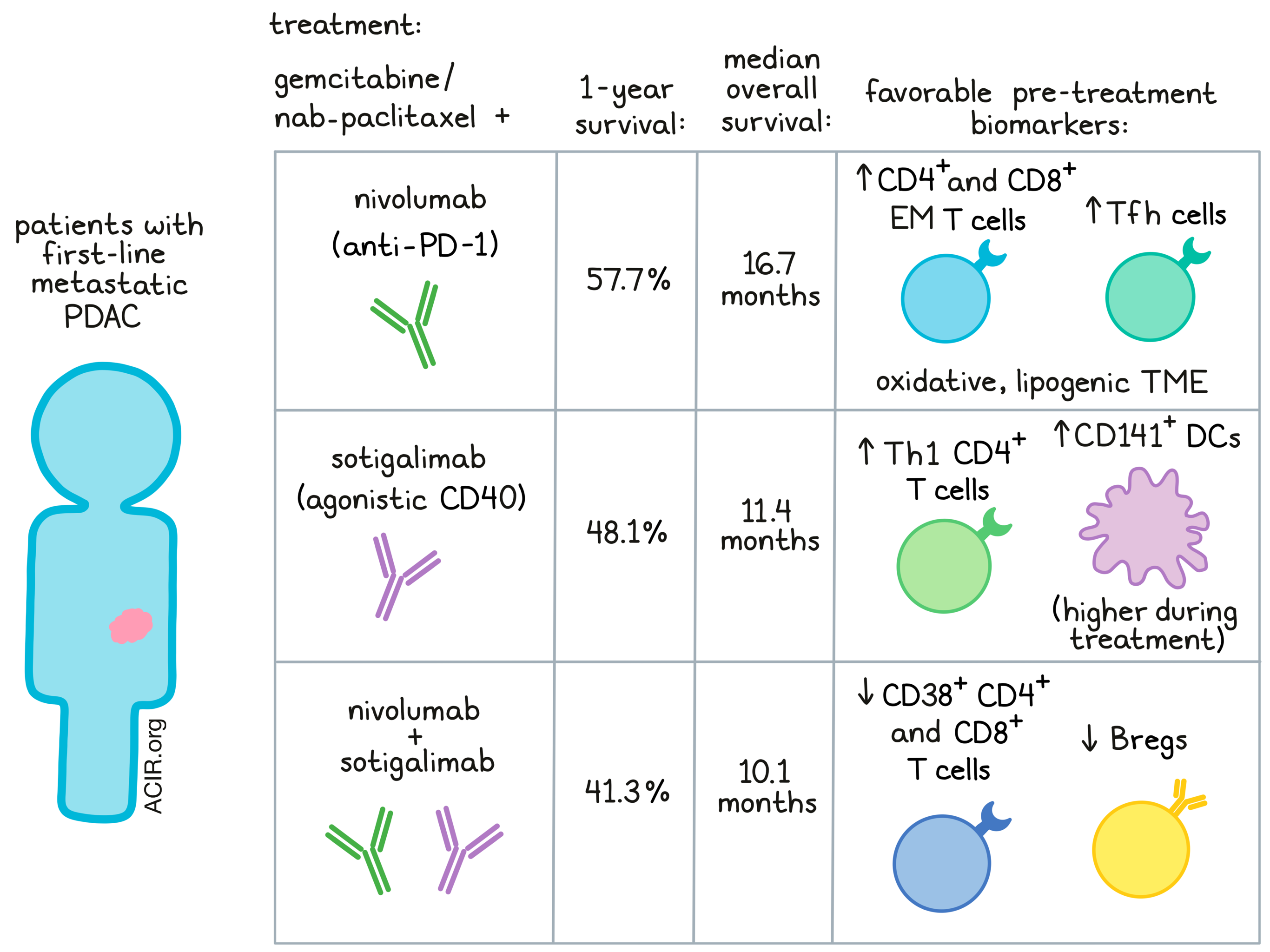
Only the nivo/chemo arm met the primary endpoint, improvement in 1-year OS rate compared to a historical chemotherapy cohort, with 57.7% versus 35% OS and a 16.7-month median OS. The sotiga/chemo and sotiga/nivo/chemo groups had 1-year OS rates and median OS of 48.1%/11.4 months and 41.3%/10.1 months, respectively; these results were not significantly different from the control group. Treatment-related adverse events occurred in almost all patients (98%) and were consistent among groups. Notably, the nivo/chemo group had the lowest rate of cytokine release syndrome (0% nivo/chemo vs. 24% sotiga/chemo and 34% sotiga/nivo/chemo; most CRS events were below Grade 3, and there were no Grade 4 or 5 events), although a higher proportion of patients in this group discontinued study due to adverse events.

Importantly, the researchers interrogated systemic and tumor features following treatment, with the goal of identifying biomarkers for treatment efficacy. RNA sequencing, CYTOF, multiplex IHC, and serum protein profiling were employed. Broadly, all treatment groups increased proliferation and activation of circulating T cells, along with serum cytokine (e.g., IFNγ) and chemokine (e.g., CXCL9, CXCL10) levels. Nivo-containing groups decreased PD-L1 tumor expression over treatment.

The team next turned to specific biomarkers associated with OS in each treatment arm. In nivo/chemo patients, pretreatment blood frequencies of effector memory CD8+ T cells expressing PD-1 and Tbet and antigen-experienced (PD-1+CD39+) effector memory CD4+ T cells expressing CTLA-4 and ICOS associated with enhanced survival. Notably, on-treatment circulating T follicular helper cell frequencies correlated most strongly with improved survival in this arm. In the tumor microenvironment, multiple gene expression signatures were related to survival outcomes, including metabolic pathways. For example, an oxidative, lipogenic tumor microenvironment was associated with better patient survival. On the other hand, gene signatures for TNFα, TGFβ, and NFκB signaling linked to poorer survival.

Interestingly, the biomarkers predictive of survival in the sotiga/chemo group diverged significantly from those found in nivo/chemo response. Here, Padrón, Maurer, and O’Hara especially considered antigen-presenting cell markers, based on CD40’s known mechanism of action and earlier findings from mouse models. In the sotiga/chemo arm, higher pretreatment frequency of cross-presenting (CD1c+CD141+) DCs, B cells, and antigen-experienced CD4+ T cells in blood related to improved survival. Similarly, on-treatment frequencies of circulating CD141+ DCs, conventional DCs, and Th1 CD4+ T cells were associated with better survival outcomes. In the tumor, CD4+ T cell gene signatures, particularly expressing Th2, Th1, and IFNγ, connected to improved survival.

Although the blood and tumor biomarkers relating to treatment efficacy largely differed between the nivo/chemo and sotiga/chemo groups, circulating IL-6 and tumor-expressed TGFβ aligned with reduced survival in both. However, the authors found little overlap between either of these groups’ biomarkers and those of the sotiga/nivo/chemo arm. In patients treated with sotiga/nivo/chemo, specific biomarkers associated with longer survival included lower frequencies of activated (CD38+) non-naive CD4+ and CD8+ T cells and CCR7+CD11b+CD27- B cells – potentially a regulatory B cell phenotype. Interestingly, circulating CD8+ T cells did not appear positively associated with response in any arm, perhaps pointing to a unique role of CD4+ T cells in the PDAC TME.

In summary, this trial and the extensive correlative data suggest that combination chemo-immunotherapy may provide value in treating PDAC, showcasing improved clinical responses and extended OS compared to a chemo-only cohort. The authors postulate that a particular treatment combination may not be appropriate for all PDAC patients; rather, the pre-treatment biomarkers identified here could lend relevance to one approach over the others, potentially attributable to the different mechanisms of action of these therapies. Additional patient trials combining response data with local and systemic immune characterization may continue to shed light on optimal treatment combinations and accelerate therapeutic development for refractory diseases such as PDAC.

Write-up by Alex Najibi, image by Lauren Hitchings

References:

Padrón LJ, Maurer DM, O'Hara MH, O'Reilly EM, Wolff RA, Wainberg ZA, Ko AH, Fisher G, Rahma O, Lyman JP, Cabanski CR, Yu JX, Pfeiffer SM, Spasic M, Xu J, Gherardini PF, Karakunnel J, Mick R, Alanio C, Byrne KT, Hollmann TJ, Moore JS, Jones DD, Tognetti M, Chen RO, Yang X, Salvador L, Wherry EJ, Dugan U, O'Donnell-Tormey J, Butterfield LH, Hubbard-Lucey VM, Ibrahim R, Fairchild J, Bucktrout S, LaVallee TM, Vonderheide RH. Sotigalimab and/or nivolumab with chemotherapy in first-line metastatic pancreatic cancer: clinical and immunologic analyses from the randomized phase 2 PRINCE trial. Nat Med. 2022 Jun 3. 

In the Spotlight...

An innate IL-25-ILC2-MDSC axis creates a cancer-permissive microenvironment for Apc mutation-driven intestinal tumorigenesis

Jou et al. demonstrated that IL-25-activated group 2 innate lymphoid cells (ILC2) created an immune suppressive microenvironment and promoted mApc-driven intestinal tumorigenesis. Higher levels of IL-25 were associated with poor prognosis in human intestinal tumors, and genetic ablation of IL-25 reduced intratumoral ILC2s and MDSCs, decreased tumor growth, and improved survival of Apc1322T/+ spontaneous mouse model of CRC. ILC2-produced IL-4 and IL-13 promoted MDSC-mediated immune suppression. Therapeutic blockade of the IL-25–ILC2 axis reduced intratumoral ILC2s and MDSCs, enhanced antitumor immunity, and decreased tumor burden.

Contributed by Shishir Pant

Fundamental immune-oncogenicity trade-offs define driver mutation fitness

To identify and quantify the factors leading to the occurrence of oncogene hotspot mutations, Hoyos and Zappasodi et al. used tp53, a critical transcription factor involved in genome integrity, as a model. Hypothesizing that the determinants were multi-factorial, a “free fitness” model was developed based on mutation rate, normalized tp53 protein concentration, a yeast assay for functional activity, and an immunogenicity score based on binding prediction. The model highlighted the known recurrent mutations and predicted a stronger role for function and weaker role for immune fitness in germline precancerous lesions, suggesting an opportunity for prophylactic immunotherapy.

Contributed by Ed Fritsch

PGE2-EP2/EP4 signaling elicits immunosuppression by driving the mregDC-Treg axis in inflammatory tumor microenvironment

Thumkeo and Punyawatthananukool et al. demonstrated that PGE2-EP2/EP4 signaling promoted active inflammation through NF-κB-mediated upregulation of target proinflammatory and angiogenesis genes in myeloid cells and simultaneously induced immunosuppression through mregDC-mediated Ccl22 and Ccl17 production to facilitate Treg recruitment and activation in the tumor. EP2/EP4 inhibition reduced NF-κB dependent gene expression, decreased tumor-infiltrating Tregs, induced type I IFN gene signatures, and suppressed LLC1 tumor growth. Expression of PTGER2 and PTGER4 (encoding EP2 and EP4) together correlated with poor prognosis in several cancers.

Contributed by Shishir Pant

Single-cell RNA-seq-based proteogenomics identifies glioblastoma-specific transposable elements encoding HLA-I-presented peptides

Using single-cell and bulk transcriptomic samples and immunopeptidomic datasets, Bonté and Arribas et al. identified HLA-I-bound peptides encoded by transposable elements (TEs) that were differentially expressed in glioblastoma (GBM) and were immunogenic in vitro. Most peptides were encoded by repeat sequences from intact ORFs that were redundant across several hundred younger TEs, while some were encoded by single copies of TEs from old subfamilies. The latter were more likely to be recurrently and differentially expressed in GBM, and could serve as strong targets for immunotherapy.

Contributed by Lauren Hitchings

cDC1 vaccines drive tumor rejection by direct presentation independently of host cDC1

Ferris et al. showed that GM-CSF-derived DCs (GMDCs) and Flt3L-derived cDC1s, but not cDC2s, cross-presented cell-associated antigens to CD8+ T cells in vitro. In contrast, in Irf8 +32-/- mice, which lack host cDC1s and are unresponsive to subcutaneous (s.c.) immunogenic fibrosarcomas, intratumoral (i.t.), but not intravenous (i.v.) injection of in vitro-generated cDC1s, but not GMDCs or cDC2s, induced rejection of the injected and uninjected contralateral s.c. tumors. The i.t.-injected cDC1s migrated to tumor-dLNs and stimulated tumor-specific CD4+ T and CD8+ T cells to induce primary and recall tumor rejection. Ex vivo antigen loading of cDC1s was not required.

Contributed by Paula Hochman

MHC class II molecules on pancreatic cancer cells indicate a potential for neo-antigen-based immunotherapy

Baleeiro et al. studied the role of ectopically expressed MHC class II, found on over 75% of pancreatic ductal adenocarcinoma (PDAC) tumors and on several PDAC cell lines. In contrast to having an immune evasive role, the MHC-II/LAG3 pathway enhanced CD8+ and CD4+ cytotoxicity towards MHC-II-positive PDAC cells, but not proliferation or IFNγ production. In silico analysis from 127 PDAC tumors revealed a number of neoantigens that could be presented by patients’ HLA class II alleles and were immunogenic in vitro, suggesting that PDAC cells display functional MHC-II molecules loaded with tumor-derived peptides that could be harnessed for immunotherapy.

Contributed by Katherine Turner

Everything New this Week In...

Close Modal

Small change for you. Big change for us!

This Thanksgiving season, show your support for cancer research by donating your change.

In less than a minute, link your credit card with our partner RoundUp App.

Every purchase you make with that card will be rounded up and the change will be donated to ACIR.

All transactions are securely made through Stripe.