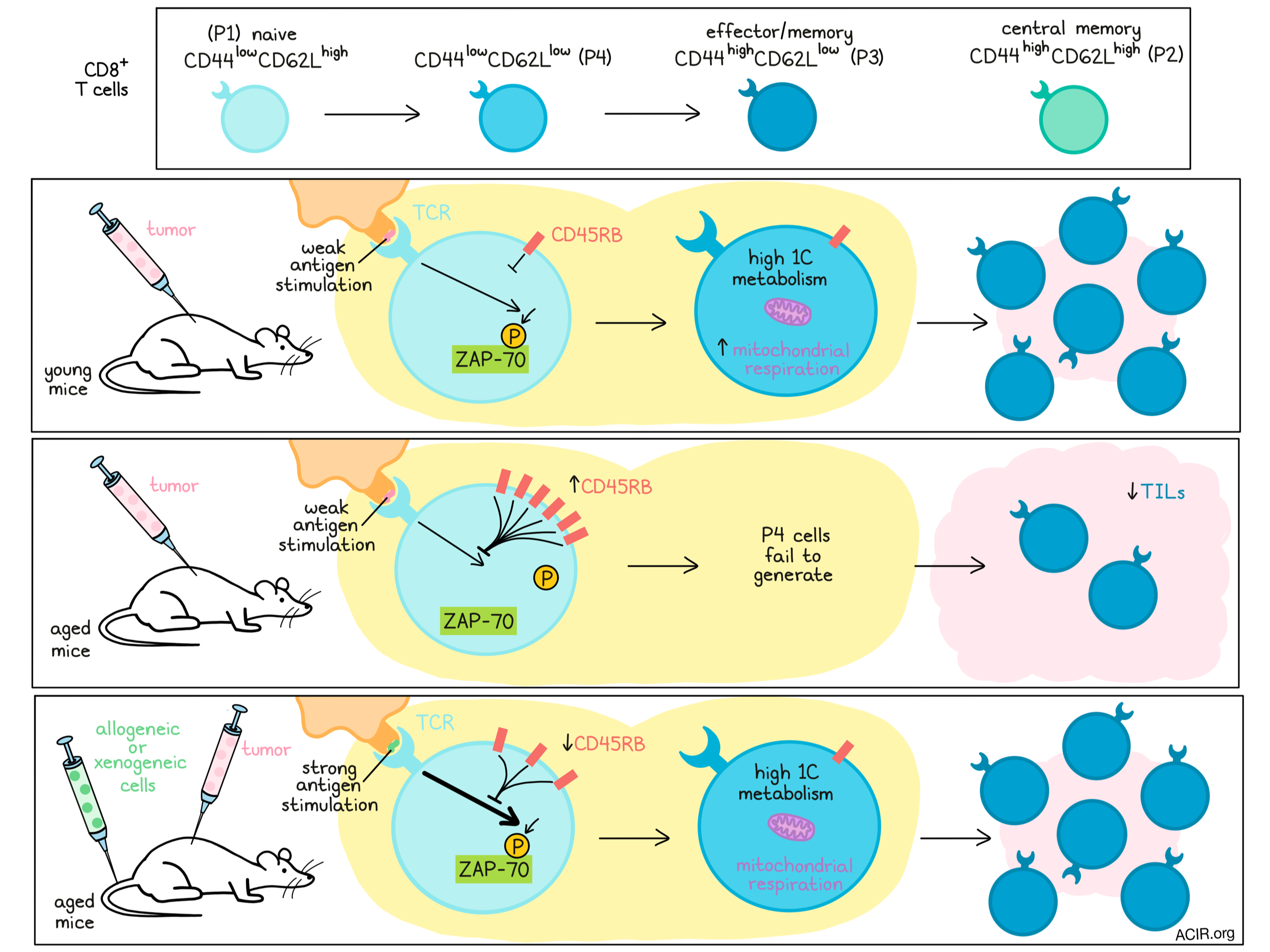
With advancing age comes a decrease in immune function, which can also dampen responses to immunotherapy. Investigating this, Nakajima et al. compared T cell subsets, based on their expression of CD44 and CD62L, in young versus aged mice. Among CD8+ T cells, CD44lowCD62Lhigh (P1) cells are defined as naive, CD44highCD62Lhigh (P2) cells are defined as central memory, and CD44highCD62Llow (P3) cells are defined as effector memory. Interestingly, CD44lowCD62Llow (P4) cells, which are rare and poorly defined, expanded after tumor inoculation in young, but not old mice, prompting further investigation into this subset. The findings were recently published in PNAS.
Using PD-1-knockout and wild-type mouse models, Nakajima et al. showed that aged mice failed to mount strong antitumor immunity and failed to generate P4 cells upon tumor inoculation. Looking more closely into this cell subset, they found that P4 cells had the potential for antitumor activity, as adoptive transfer of P4 cells controlled tumors as well as adoptive transfer of P3 cells. By day 6 after transfer, all of the transferred P4 cells had differentiated into P3 cells, suggesting that P4 cells may mediate antitumor immunity as precursors to effector/memory cells. Ex vivo stimulation studies showed that P4 cells differentiated from P1, but not P2 cells.
Looking more closely at gene expression in the P4 subset, the researchers found that genes related to activation were higher in P4 cells than in P1 cells, and genes associated with differentiation were lower in P4 cells than in P3 cells, further supporting the notion that P4 cells are pre-effector-like cells in an early activation state. Next, gene ontology enrichment analysis revealed that genes for enzymes associated with 1 carbon (1C) metabolism (a set of biochemical reactions that provide single carbon methyl units for multiple biosynthetic pathways) were uniquely upregulated in P4 cells. Genes for 1C metabolism were enriched upon tumor inoculation in young, but not aged mice, in line with previous data on the generation of the P4 subset. This was further accompanied by an increase in mitochondrial function. Consistent with this, T cells from aged tumor-bearing mice showed overall lower rates of oxygen consumption compared to their younger counterparts.
To better understand why P1 cells fail to differentiate into P4 cells in aged mice, Nakajima et al. found that TCR signaling was attenuated in P1 cells from older mice, as evidenced by limited ZAP-70 phosphorylation. After showing that TCR expression levels and avidity were unchanged, the researchers turned their attention towards CD45RB (the major CD45 isoform in naive and memory T cells in mice), which, when present at a high density on the cell surface, can weaken TCR signaling in naive T cells. In bulk CD8+ T cells from aged mice, CD45RB expression was higher, and this effect was greater when looking specifically at the P1 and P4 subsets. When CD45RB was blocked using a CD45 inhibitor, ZAP-70 phosphorylation increased in both aged and young mice, suggesting that CD45RB suppresses CD8+ T cell activation and differentiation from P1 to P4 by interfering with TCR signal transduction.
Interestingly, when P1 cells from aged mice were stimulated ex vivo using anti-CD3/CD28 mAbs, they differentiated into P4 and P3 cells as effectively as P1 cells from young mice. This suggested that while nominal antigenic stimulation was insufficient to generate P4 cells, strong TCR stimulation could overcome this deficit.
In an effort to restore differentiation from P1 to P4 cells in aged mice, the researchers introduced xenogeneic cells (Daudi cells) to induce strong antigenic stimulation. Following injection of Daudi cells, the researchers observed increased P4 cells, increased 1C metabolism, and reduced CD45RB in P1 cells in both young and aged PD-1-knockout mice. When aged PD-1-knockout mice treated with Daudi cells were inoculated with MC38 tumors, they showed decreased tumor growth and increased survival compared to untreated mice. In wild-type mice, age-related unresponsiveness to PD-1 blockade was also recovered with Daudi cell injection, or with injection of splenocytes from Balb/c mice, which also served as effective alloantigens.
Looking more closely, the researchers found that Daudi cells increased phosphorylation of ZAP-70 in T cells, and increased the proliferation of tumor antigen-reactive CD8+ T cells. Within tumors, most CD8+ T cells were already differentiated into P3 cells, suggesting that strong antigen stimulation enhances the differentiation of antigen-specific T cells from naive to effector, thus increasing effector T cell infiltration into tumors.
Investigating the potential of using Daudi cells in a therapeutic setting, the researchers tested whether administration of Daudi cells could restore antitumor immunity in mice with established tumors. In this setting, Daudi cells increased P4 cells, TILs, and ZAP-70 phosphorylation. These results suggest that strong immune stimulation by xenogeneic or allogeneic cell transplant could potentially recover TCR signal transduction and P4 generation to rescue compromised antitumor immunity and responses to PD-1 checkpoint blockade in aged mice.
Overall, this research highlights the importance of CD44lowCD62Llow (P4) CD8+ T cells as an intermediate cell type between naive (P1) and effector/memory (P3) CD8+ T cells. The P4 subset expresses genes associated with 1C metabolism and mediates enhanced mitochondrial function that is required for sufficient T cell activation. In aged mice, accumulation of CD45RB in P1 cells attenuates TCR signaling and limits the formation of the P4 subset. Nakajima et al. showed that this limitation can be overcome by CD45 inhibition or strong antigenic stimulation through the introduction of xenogeneic or allogeneic cells. This work provides important insights that could lead to the development of new targets to enhance antitumor immunity and restore responses to checkpoint blockade in older patients, who are often less likely to reap the benefits of immunotherapy.
By Lauren Hitchings
Meet the researcher
This week, first author Yuka Nakajima answered our questions.
What prompted you to tackle this research question?
Despite clinical studies showing a remarkable efficacy of cancer immunotherapy by PD-1 blockade, many cancer patients cannot respond to the therapy. Further, while previous work has given us information about the linkage between aging and impaired immune responses, little is known about how aging affects the efficacy of PD-1 blockade therapy. Actually, aged PD-1 knockout mice showed strong resistance to tumor growth. Therefore, we tried to better understand the mechanism using the mouse model for PD-1 blockade therapy to overcome the resistance.
What was the most surprising finding of this study for you?
The most remarkable finding was the observation that the reduction of CD44lowCD62Llow cell frequency could be correlated with resistance to PD-1 blockade therapy in aged mice. In mouse CD8+ T cells, the CD44lowCD62Llow subset had been known as a remaining minor subset, an exception to the naive, effector, and memory subsets, and the importance was not known. Normally, it is difficult to detect the CD44lowCD62Llow cells because of the transient induction from naive cells in response to TCR stimulation. Luckily, we could use PD-1 KO mice that can strongly induce TCR-dependent T cell activation. Therefore, we could easily detect CD44lowCD62Llow cells in those mice. This finding helped us better understand how aging modulates antitumor immunity through CD8+ T cell differentiation.
What was the coolest thing you’ve learned (about) recently outside of work?
Recently, I found a very beautiful cherry blossom area, when I took a walk for a change of pace during telework. Although the area was very close to my house, I didn’t know it at all. Due to the COVID-19 pandemic, we have to change our lifestyle to adapt to the new normal. Under the circonstances, I realized, “in the middle of difficulty lies opportunity”!