While immunotherapies work great in some patients, for most, results are limited. One reason for the limited therapeutic response is the induction of an “exhausted” phenotype in CD8+ TILs. Aiming to reinvigorate exhausted CD8+ TILs in the TME, Guo and Xie et al. explored the effects of IL-10 treatment and combined it with adoptive cell transfer (ACT) and checkpoint blockade in various animal models; their data were recently published in Nature Immunology.
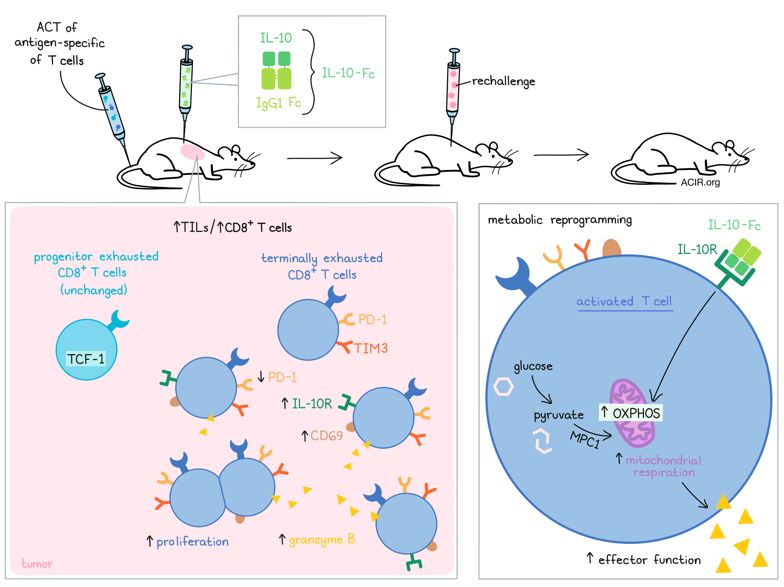
The researchers produced a recombinant fusion protein of human IL-10 and IgG1 Fc (IL-10-Fc), which has an extended half-life and cross-reacts with the mouse IL-10 receptor (IL-10R). To test its effect on reinvigorating terminally exhausted CD8+ TILs, mice with B16F10 tumors were treated with intravenous ACT of PMEL CD8+ T cells recognizing the gp100 tumor antigen, and with peritumoral administration of IL-10-Fc. The ACT increased TIL counts, which were further elevated when IL-10-Fc was coadministered, with a particular rise in CD8+ TILs.
To assess which CD8+ TIL subset increased during treatment, the researchers assessed the TILs based on their expression of TCF-1, TIM3, and PD-1. Terminally exhausted TCF-1-TIM3+CD8+ TIL increased in the tumor, while TCF-1+TIM3-CD8+ progenitor-exhausted TIL were unchanged after treatment. The TCF-1-TIM3+ population overlapped with the PD-1+TIM3+ subset, so PD-1+ and TIM3+ were used in further experiments to analyze the terminally exhausted population. The PD-1+TIM3+ cells also had increased BrdU incorporation and Ki67 expression, indicative of increased proliferation capacity. In addition, the expanded subsets produced granzyme B and other effector molecules, and expressed higher levels of IL-10R and CD69 and lower levels of PD-1.
To assess whether the effects of IL-10-Fc were antigen-dependent, the researchers performed a cotransfer experiment in the B16F10 model with antigen-specific PMEL T cells and non-specific OT-1 T cells, and found that only the PMEL cells expanded. The PD-1+TIM3+CD8+ T cells expanded even when PD-1+TIM3- progenitor exhausted T cells were excluded from the infusion. Ex vivo cultures and in vivo experiments with the progenitor T cell population (TCF-1+) depleted confirmed that the terminally exhausted cells expanded, and that increases were not the result of progenitor-exhausted cells becoming terminally exhausted.
To assess the effects of IL-10-Fc on CD8+ T cell metabolism, CD8+ T cells were TCR-stimulated with peptide-pulsed APCs or a dimerized CD3 antibody in vitro. Naive and primed CD8+ T cells upregulated OXPHOS when treated with IL-10-Fc. To mimic chronic stimulation in the tumor microenvironment, activated PMEL CD8+ T cells were co-cultured with B16F10 cells. Again, OXPHOS was upregulated after IL-10-Fc treatment. These effects were not found when IL-10Rα-knockout CD8+ T cells were used. Therefore, IL-10-Fc treatment may reprogram T cell metabolism toward OXPHOS dependence.
Guo and Xie et al. then moved to human CAR T cells directed at HER2 and co-cultured these cells with the HER2-expressing ME275 melanoma and SKOV3 ovarian cancer cell lines. The CAR T cells also upregulated OXPHOS and had enhanced proliferation and cytotoxicity when co-treated with IL-10-Fc. Additionally, overstimulation of human CD8+ T cells resulted in a PD-1+LAG3+ population with high expression of IL-10Rα, and these cells responded similarly to IL-10-Fc treatment as mouse PD-1+TIM3+CD8+ T cells.
Moving to assess the antitumor effects of treatment, the researchers administered ACT and IL-10-Fc to the B16F10 model. This treatment regimen resulted in durable tumor eradication in 90% of mice, while ACT alone only transiently controlled tumor growth, and IL-10-Fc alone eradicated tumors in only 30% of mice. The combination strategy resulted in high infiltration of cytotoxic and polyfunctional T cells. Of the survivors, ~80% rejected a tumor rechallenge, indicating immune memory. These results were confirmed in an OVA-expressing YUMM1.7 melanoma model treated when there was a large, established tumor. In this model, the combination strategy also resulted in durable responses, with 60% of mice surviving long-term and rejecting a rechallenge.
To test the combination of IL-10-Fc and CAR T cell therapy, mice were inoculated with MC38-HER2 colon adenocarcinoma and were treated with HER2-CAR-T and IL-10-Fc. The CAR T cells alone had little effect on the tumor, but the combination eradicated tumors and led to long-term survival in 90% of mice, which all rejected a rechallenge. Finally, the researchers evaluated the combination of IL-10-Fc and anti-PD-1 antibodies in a mouse CT26 colorectal model. The combination treatment resulted in 60% survival, and over 80% of surviving mice also rejected a rechallenge.
Diving deeper into the mechanistic changes in the terminally exhausted CD8+ TIL after treatment, the researchers performed RNAseq on sorted antigen-specific Thy1.1+PD-1+TIM3+CD8+ TILs from B16F10 tumors that were treated with ACT and IL-10-Fc. The PD-1+TIM3+CD8+ T cells had higher expression of genes associated with the electron transport chain, which is OXPHOS-related, and genes encoding cytotoxic molecules. Meanwhile, genes encoding inhibitory receptors and exhaustion transcription factors were downregulated. In addition, gene set enrichment and pathway analysis revealed enrichment of signatures related to T cell OXPHOS and effector function. Furthermore, mitochondrial profile analysis revealed that mitochondrial respiration was upregulated in PD-1+TIM3+CD8+ TILs.
Using a set of pathway-specific inhibitors, Guo and Xie et al. found that the enhanced OXPHOS upon IL-10-Fc treatment was not due to increased fatty acid oxidation or glutaminolysis, but might depend on pyruvate generated from glycolysis. This was confirmed using OT-1 cells deficient in the Mitochondrial Pyruvate Carrier 1 (MPC1) gene. These OT-1 cells did not upregulate OXPHOS, enhance mitochondrial function, or expand upon IL-10-Fc treatment. Confirming these data in vivo, MPC1-knockout OT-1 cells did not expand, and treatment had reduced efficacy in two mouse models.
The data in this report suggest that combining immunotherapies with IL-10-Fc can reinvigorate terminally exhausted T cells and enhance the efficacy of immunotherapy. If these data are confirmed in patients, this new strategy may increase the number of patients benefiting from immunotherapy. Further work could also lead to metabolic therapies that directly stimulate OXPHOS in the ever-present, but ineffective exhausted T cells.
Write-up by Maartje Wouters, image by Lauren Hitchings
Meet the researcher
This week, first author Yugang Guo answered our questions.
What prompted you to tackle this research question?
Cancer immunotherapies, represented by immune checkpoint blockades and adoptive T cell transfer therapy, have achieved remarkable clinical success. However, an outstanding challenge remains that a great majority of patients still can’t benefit from these expansive therapies, partially because of T cell exhaustion. Interleukin-10 (IL-10) is a pleiotropic cytokine, which promotes antitumor immunity in multiple murine tumor models. However, the mechanism of how IL-10 could boost the exhausted T cell response is still largely unknown. Given the sense that T cell metabolism determines T cell fate, we were very interested to investigate whether IL-10 could also reprogram T cell metabolic profiles and restore the function of exhausted T cells, particularly in terminally exhausted T cells, which are currently believed to be difficult to be reactivated by most existing immunotherapies, including checkpoint blockades.
What was the most surprising finding of this study for you?
Among the exhausted tumor-infiltrating T cells, a subset called “terminally exhausted CD8+ T cell”, does not respond to most existing immunotherapies, including immune checkpoint blockades. Our study shows that metabolic reprogramming of these cells by IL-10-Fc, through a molecule called mitochondrial pyruvate carrier (MPC), is sufficient to restore the functionality of terminally exhausted CD8+ T cells for fighting cancer. This finding lays the foundation for further identification of metabolic stimulations that are needed for reinvigorating terminally exhausted CD8+ T cells, a current major bottleneck in the field of cancer immunotherapy. Furthermore, we were really surprised that metabolic intervention on terminally exhausted T cells significantly unleashed the potential of cancer immunotherapy (especially in the context of adoptive T cell therapy) against solid cancers, leading to eradication of established solid tumors and durable cures in a majority of treated mice.
What was the coolest thing you’ve learned (about) recently outside of work?
I like to go jogging around the Leman lake outside of work. I usually run 8 kilometers in 50 minutes each time, and have run more than 2,400 kilometers in total during the past 2 years (even in the difficult time of COVID-19 pandemic). I enjoy jogging because I feel a refreshing of the mind after running, and I also get a good sleep, which further helps me to recover from busy work. To some extent, like IL-10, jogging keeps the metabolic fitness of our body from exhaustion induced by extensive labor and mental work in daily life.