Weekly Digests
‹ Back to June

Liver cancer, immunity, and the microbiome: connecting the dots

June 13, 2018

The gut microbiome has recently emerged as a critical regulator of antitumor immunity. Nearby, the liver is supplied by blood largely from the intestines, meaning that it is directly exposed to the runoff of metabolites and other products of the microbiome. Noting this direct physical relationship, Ma et al. set out to determine whether changes in the microbiome might affect hepatic immune cells and antitumor immunity in the liver.

To begin, the researchers induced spontaneous hepatocellular carcinoma (HCC) in mice and treated them with an antibiotic cocktail (ABX) to deplete commensal gut bacteria. Surprisingly, they found fewer and smaller HCC tumors in mice treated with the antibiotics. Similarly, in liver metastasis models (subcutaneous EL4 thymoma and intrasplenic tumor injection of B16 melanoma), fewer spontaneous metastases formed in the liver when mice were treated with antibiotics, while no effects were observed on primary tumors located outside of the liver, nor on the formation of metastases in other locations, indicating a liver-specific antitumor effect.

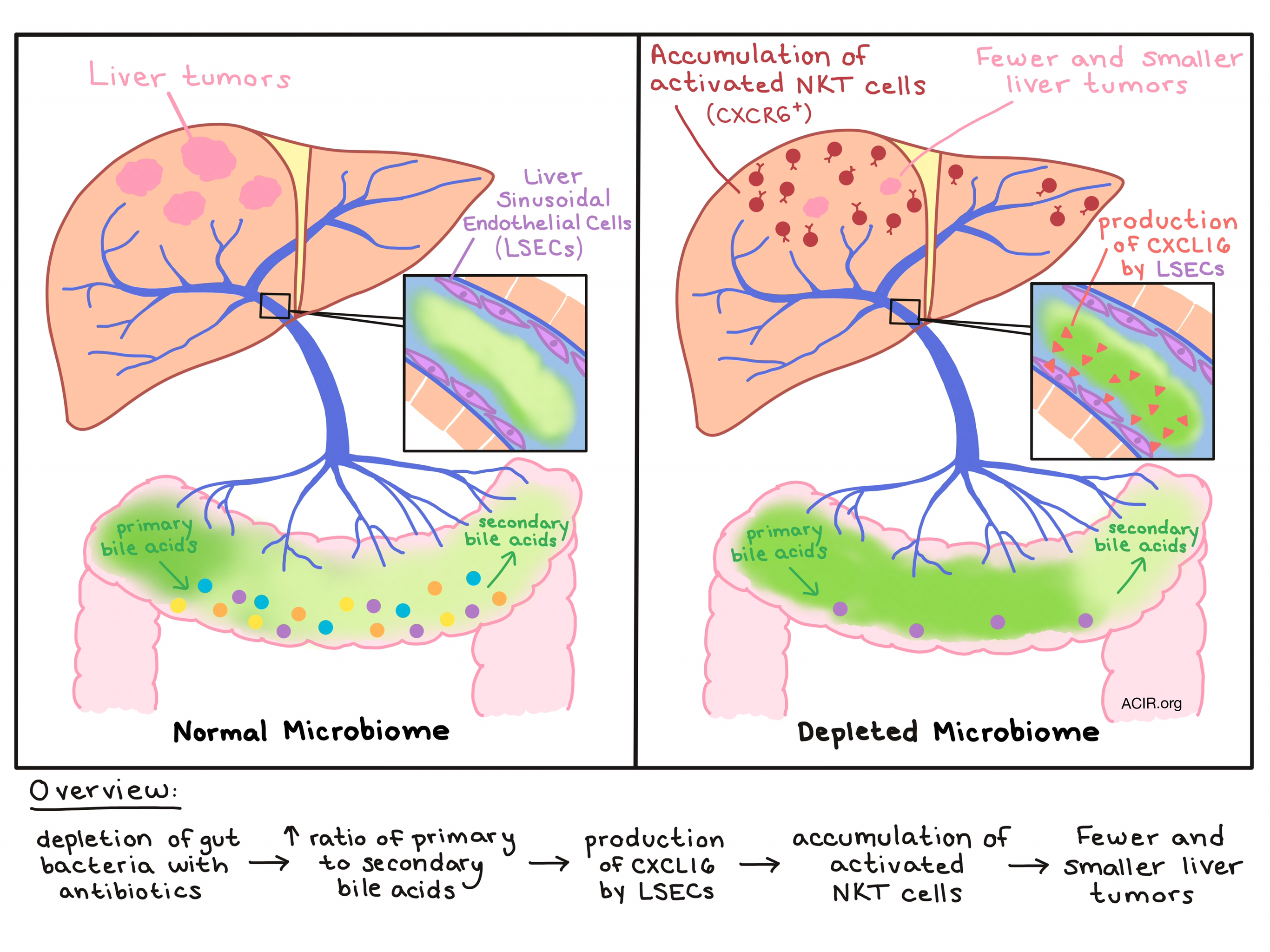
Exploring which immune cell subsets might be behind the observed antitumor effect, Ma et al. noted prominent expansion and accumulation of hepatic natural killer T (NKT) cells in the livers of ABX-treated mice, and that those NKT cells were highly activated and produced higher levels of IFNγ, both in tumor-bearing and tumor-free mice. Antibody depletion and gene knockout experiments in tumor-bearing mice showed that NKT cells were the primary mediators of tumor inhibition associated with ABX treatment.

Nearly all hepatic NKT cells express the receptor CXCR6, which interacts only with the ligand CXCL16, so unsurprisingly, Ma et al. found that ABX treatment increased mRNA levels of Cxcl16. Previously reported to be a major source of CXCL16, liver sinusoidal endothelial cells (LSECs) were found to upregulate CXCL16 production in ABX-treated mice, thus recruiting NKT cells to the liver.

Based on a known role for the cell surface-bound form of CXCL16 in lipid metabolism, Ma et al. suspected a link between CXCL16 upregulation by LSECs and bile acid metabolism mediated by commensal gut bacteria. To prove this connection, the team used a bile acid sequestrant to reduce bile acid levels in the liver and saw increased NKT cell accumulation. Digging deeper into which bile acids were at play, the researchers found that ABX treatment increased primary acids, while reducing secondary bile acids, consistent with the understanding that commensal gut bacteria convert primary bile acids into secondary bile acids. In vitro, secondary bile acids were found to suppress expression of Cxcl16 mRNA by LSECs, while primary bile acid induced expression of Cxcl16. In vivo, oral gavage of secondary bile acids reversed the ABX-mediated accumulation of NKT cells and antitumor effect in the liver, while primary bile acids had the opposite effect - further enhancing NKT cell accumulation and tumor inhibition.

By treating mice with individual antibiotics rather than a cocktail, the research team was able to identify gram positive bacteria of the Clostridium genus as having a negative effect on hepatic NKT cell accumulation and liver tumor inhibition, and reconstitution with C. scindens rapidly reduced the effects of ABX treatment. Depleting these bile-acid converting commensal gut bacteria raises the ratio of primary to secondary bile acids, inducing production of CXCL16 by LSECs in the liver. Increased CXCL16 production leads to the accumulation of activated CXCR6+ NKT cells in the liver, which can exert an antitumor effect on primary liver tumors and liver metastases in mice.

Ma et al. extended their outlook to human samples to determine whether their findings in mice might translate. Similar to what they saw in mice, primary bile acids induced CXCL16 mRNA expression in SK-HEP1 cells, representative of LSECs. Analysis of primary/secondary bile acid ratio and CXCL16 mRNA levels in data from a prior cohort of patients suggested that the opposing effects of primary and secondary bile acids on CXCL16 expression hold true in humans. In addition to providing insight into the connection between the microbiome, the immune system, and liver cancer, the results of this study may influence the design of future immunotherapies related to the treatment of liver cancer or liver metastases.

by Lauren Hitchings

References:

Ma C., Han M., Heinrich B., Fu Q., Zhang Q., Sandhu M., Agdashian D., Terabe M., Berzofsky J.A., Fako V., Ritz T., Longerich T., Theriot C.M., McCulloch J.A., Roy S., Yuan W., Thovarai V., Sen S.K., Ruchirawat M., Korangy F., Wang X.W., Trinchieri G., Greten T.F. Gut microbiome-mediated bile acid metabolism regulates liver cancer via NKT cells. Science. 2018 May 25.

In the Spotlight...

Atezolizumab for First-Line Treatment of Metastatic Nonsquamous NSCLC

In a phase 3 clinical trial, Socinski et al. compared the combination of the PD-L1 inhibitor atezolizumab, the VEGF inhibitor bevacizumab, and chemotherapy (ABCP) to bevacizumab and chemotherapy (BCP) in nonsquamous non-small-cell lung cancer. ABCP patients had longer median progression-free survival (PFS), doubled rates of PFS at 12 months, and higher overall survival than BCP patients. Prolonged PFS was improved in all subgroups (PD-L1 level, mutation status [wild type or EGFR/ALK mutant], or Teff gene expression). The safety profile was consistent with the known profile of the agents.

Pharmacologic inhibition of protein phosphatase-2A achieves durable immune-mediated antitumor activity when combined with PD-1 blockade

Ho et al. found that the small molecule LB-100, an inhibitor of protein phosphatase-2A (PP2A), in combination with anti-PD-1, induced CD8+ T cell-dependent tumor regression in a majority (complete response in half) of mice with established CT26 colorectal tumors, and protected complete responders from rechallenge. Mechanistically, the combination increased tumor infiltration by CD8+ T cells with enhanced effector and cytolytic functions. LB-100 also activated the mTORC1 pathway, reducing CD4+ T cell differentiation into Tregs and promoting Th1 cytokine secretion. LB-100 and anti-PD-1 prophylactically controlled B16 melanoma.

Radiotherapy and CD40 activation separately augment immunity to checkpoint blockade in cancer

In a mouse model of pancreatic ductal adenocarcinoma, Rech et al. found that the combination of radiotherapy (RT), agonist CD40 (agCD40), and anti-PD-1/anti-CTLA-4 decreased both irradiated and non-irradiated tumor burden and improved survival. The antitumor effect was dependent on CD4+ and CD8+ T cells, short-lived myeloid cells, and host CD40, IFNγ, and Batf3. Random forest analysis indicated that key predictors of response were RT ablation of intratumoral CD4-/CD8- T cells, reconfiguration of myeloid compartments by agCD40, increased unirradiated intratumoral CD8+ T cells, and increased CD8+/Treg ratio at peak tumor regression.

Co-transfer of tumor-specific effector and memory CD8+ T cells enhances the efficacy of adoptive melanoma immunotherapy in a mouse model

Co-transfer of tumor-specific CD8+ Teff and Tmem cells by Contreras et al. led to the strongest control of tumor growth in a B16F10 murine model in comparison to transfer of either alone. Co-transfer produced the strongest intratumoral infiltration by endogenous CD8+ T cells and the greatest antigen-specific expression of IFNγ in splenocytes. Paracrine release of IL-2 from Tmem also appeared to increase Teff inhibition of melanoma cell proliferation, and temporal differences in ability of Tmem and Teff to inhibit proliferation may have contributed to the success of the combination.

Induction of anergic or regulatory tumor-specific CD4+ T cells in the tumor-draining lymph node

Using a genetically engineered autochthonous lung adenocarcinoma mouse model expressing a cytoplasmic neoantigen, Alonso et al. demonstrate that naive, tumor antigen (Ag)-specific CD4+ T cells become partially activated and proliferate in the tumor-draining lymph node (TdLN), but do not accumulate in the tumor regardless of Ag load or tumor size. Instead, the majority of these cells become anergic, and a small but significant fraction independently differentiate into peripherally-induced pTregs. CD4+ T cell anergy required continuous Ag presentation by the tumor and was dependent on the host Tregs present in the TdLN.

Adaptive NK cells resist regulatory T cell suppression driven by IL-37

Aiming to uncover novel approaches to utilize NK cells, Sarhan et al. found that while FcεRγ+NKG2C- canonical NK cells are susceptible to Treg suppression, FcεRγ-NKG2C+ adaptive NK cells are resistant to it. Tregs secrete IL-37, which binds the inhibitory receptor IL1R8 on canonical NK cells, exerting functional suppression by downregulating TIM-3 (which acts as an activating receptor on NK cells) and upregulating PD-1 and IL1R8. Adaptive NK cells were found to be resistant to Treg suppression due to significantly lower expression of IL1R8 compared to canonical NK cells.

Tissue-resident memory T cells at the center of immunity to solid tumors

In this comprehensive review of tissue-resident memory T (TRM) cells, Amsen et al. discuss the properties that define this recently uncovered and distinct T cell lineage, how they are generated and maintained, and their role in fighting solid tumors. Often marked by CD103 expression, TRM cells adapt to and reside in a specific tissue environment where they persist long term to protect against recurrent threats. These adaptations, along with their ability to adhere to their tissue of residence, grant TRM cells unique “home field” advantage for battling solid tumors that could one day be exploited for immunotherapy.

Tumor immune evasion arises through loss of TNF sensitivity

Using whole-genome CRISPR/Cas9 screening on MC38 colon adenocarcinoma and B16 melanoma cell lines, Kearney et al. demonstrated that tumors evade CD8+ T cell killing by disrupting antigen presentation, IFNγ, and TNF signaling, and evade NK cells by disrupting TNF signaling. Perforin-related pathways were not involved in evasion mechanisms. Recombinant TNF (but not IFNγ) was able to directly kill tumor cells and, upon antigen recognition, TNF mediated bystander killing of antigen-negative tumor cells in vitro. Analysis of patient data in TCGA confirmed these observations.

Everything New this Week In...

Close Modal

Small change for you. Big change for us!

This Thanksgiving season, show your support for cancer research by donating your change.

In less than a minute, link your credit card with our partner RoundUp App.

Every purchase you make with that card will be rounded up and the change will be donated to ACIR.

All transactions are securely made through Stripe.