Antigen cross-presentation by dendritic cells (DCs) is traditionally thought of as the primary mechanism underlying CD8+ T cell priming against tumors, but evidence of an alternative antigen presentation pathway, DC cross-dressing with tumor-derived peptide:MHC-I, has also been observed. In a study recently published in Immunity, McNabb et al. investigated the contributions of this alternative antigen presentation pathway in inducing antitumor immunity.
To begin, MacNabb et al. established a C1498 leukemia model expressing the Kb-restricted antigen SIY tagged with eGFP. Knocking out Kb eliminated tumor cell presentation of the antigens on MHC-I, but did not impact SIY expression, bone marrow-derived DC cross-presentation of SIY from tumor cell lysates, or DC-mediated proliferation of CTV-labeled SIY-specific 2C CD8+ T cells. When Kb was expressed in implanted tumor cells, 2C CD8+ T cells proliferated, accumulated, and upregulated IFNγ, TNFα, and granzyme B. However, these effects were lost when Kb-/- tumors were implanted, suggesting that Kb expression on tumor cells was required for effective T cell priming in this model. Similar studies were performed in a B16F10 melanoma model expressing eGFP-tagged OVA and Kb-restricted SIINFEKL, though this model expressed lower levels of Kb compared to the C1498 model. In this setting, knockout of Kb expression on tumor cells did not abrogate, but showed a trend toward reducing the proliferation and accumulation of OT-I cells.
Next, looking at the effect of tumor pMHC-I expression on the priming of endogenous T cell responses in mice, the researchers found that Kb knockout in C1498.SIY tumors reduced expansion of endogenous SIY-specific T cells, along with IFNγ production. To uncouple the priming from the effector functions of T cells, primed CD8+ T cells were isolated from the tumor-draining lymph nodes (tdLNs) of mice bearing Kb+/+ or Kb-/- tumors, and were transferred in equal numbers into naive mice, which were then challenged with Kb+/+ C1498.SIY tumors. The T cells that were primed in Kb+/+ tumor setting were more effective than those primed in the Kb-/- setting, which performed no better than the controls, suggesting that Kb expression on C1498 was required for T cell priming in this model. In the B16.OVA model, the expansion of endogenous OVA-specific cells was also significantly reduced (an effect stronger than that observed with exogenous monoclonal OT-I cells), though not abrogated in mice bearing Kb-/- tumors, further suggesting that Kb expression was required for optimal T cell priming.
To determine whether this effect was due to DC cross-dressing in cancer cell-derived MHC-I molecules, as hypothesized, MacNabb et al. used flow cytometry and surface staining to show that host DCs and macrophages in tumors in Kb-/- mice acquired Kb from implanted Kb+/+ C1498.SIY tumors. To show that this effect was not just due to the lack of MHC-I on host APCs, and to assess the localization of cancer-derived Kb molecules, the researchers utilized a version of the C1498.SIY model with Kb knocked out and then re-expressed marked with eGFP. When this tumor cell line was implanted into Kb-/- mice, GFP-labeled Kb from tumor cells was identified both within and on the surface of host DCs and macrophages. In Kb-/- mice harboring Kb+/+ tumors, APC internalization of tumor antigens was found to be highly correlated with presentation of tumor-derived Kb, suggesting that MHC-I cross-dressing is related to and might occur through tumor antigen uptake. Similar results were observed using the B16.OVA model, but only when tumor cells were pre-treated with IFNγ to induce sufficient levels of Kb expression.
To determine the contribution of MHC-I cross-dressing compared to conventional MHC-I cross-presentation by APCs, MacNabb et al. transplanted Kb+/+ and Kb-/- C1498.SIY tumors into wild-type, Kb-/-, or Tap1-/- mice, which are largely incapable of classical antigen cross-presentation. This showed that tumor expression of Kb was required for proper T cell activation, while antigen cross-presentation was dispensable, as priming was similar between Tap1-/- and wild-type mice.
To test this in another model, the researchers generated and validated a Wdfy4-/- mouse model that did not produce functional WDFY4 protein and was therefore defective in cDC1 cross-presentation of cell-derived antigens. While wild-type mice typically reject 1,969 sarcoma cells, these cells were able to grow progressively in Wdfy4-/- mice. In fact, they even grew more rapidly than they did in Batf3-/- mice, which largely lack cDC1s. This suggested that in this model, classical cross-priming was required for effective antitumor responses, emphasizing the variable contributions of cross-presentation and cross-dressing in different tumor settings.
Next, mice with or without WDFY4 expression were treated with CTV-labeled antigen-specific T cells, then inoculated with C1498.SIY with or without Kb expression. While minimal T cell proliferation and expansion were observed in control mice or in mice with tumors lacking Kb expression, expansion was observed in mice bearing Kb-expressing tumors. This expansion was only slightly reduced in Wdfy4-/- mice compared to Wdfy4+/+ and Wdfy4+/- mice, suggesting that cross-presentation is not the only factor that contributes to priming. The possibility that WDFY4 expression might contribute to cross-dressing was ruled out, as APCs from mice with or without Kb expression equally acquired tumor-derived Kb, regardless of whether they expressed functional WDFY4.
To determine whether the MHC-I cross-dressing observed in mice was also relevant in human tumors, the researchers co-cultured human HLA-A*02neg monocyte-derived macrophages (MDMs) with CTV-labeled human HLA-A*02pos CD19+ OCI-Ly6 lymphoma cells. Following the administration of anti-CD47 to induce phagocytosis, MDMs captured the fluorescent signal and HLA molecules from the lymphoma cells, and presented HLA molecules on their surfaces. Further, cells with the highest fluorescence signals had the highest levels of HLA expression, validating the observation in mice that uptake of antigens was associated with expression of cancer cell-derived HLA. Interestingly, some MDM cells with high fluorescence and expression of HLA also acquired surface presentation of CD19, suggesting that surface protein transfer was not unique to MHC and HLA molecules. In a xenograft model, host DCs from the tumor, but not the spleen, acquired surface expression of HLA-I derived from human lymphoma cells.
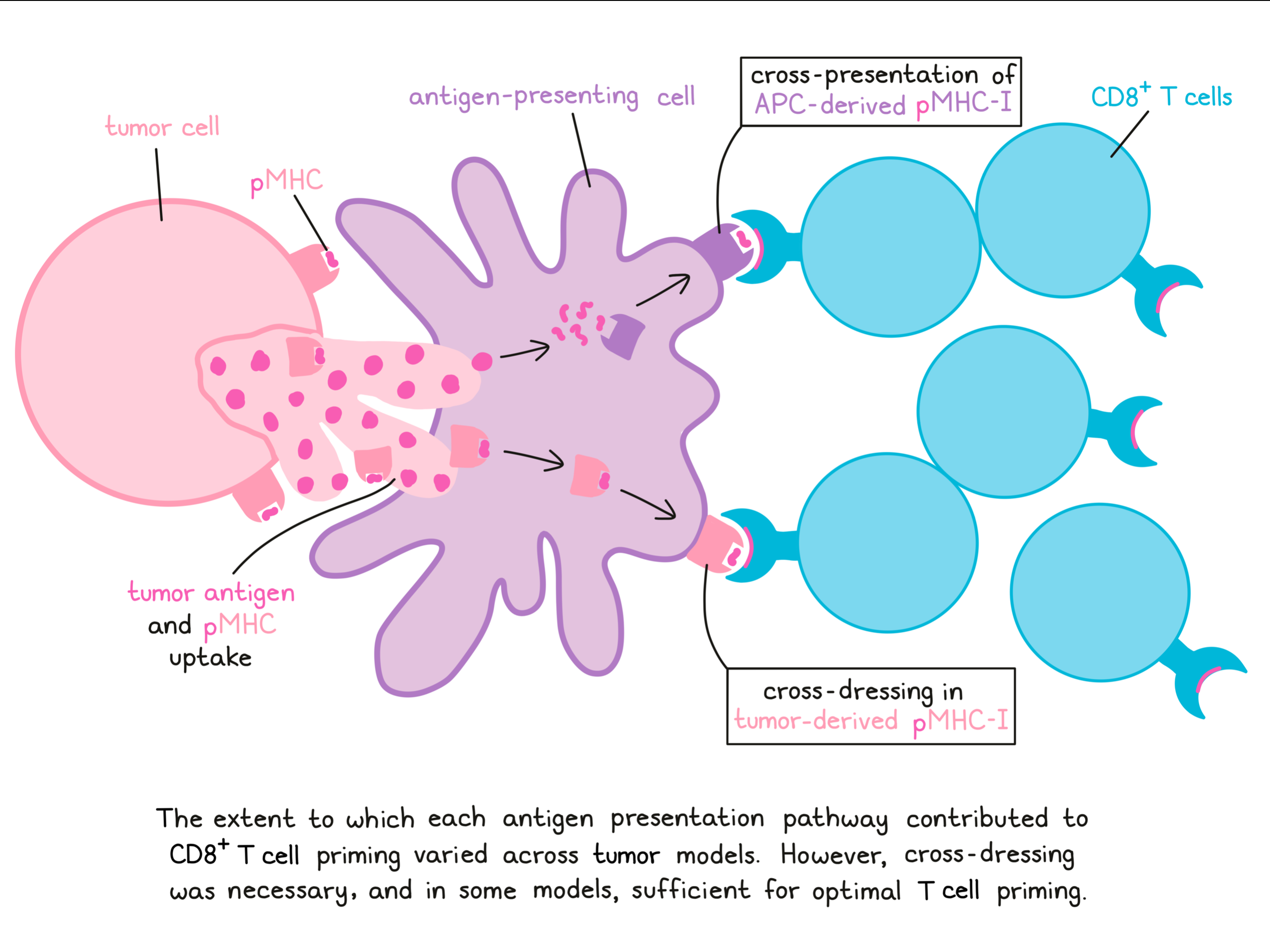
Overall, these results show that in addition to cross-presenting antigens, APCs also cross-dress in tumor-derived pMHC complexes, likely following internalization of tumor material. The contribution of this alternative antigen presentation pathway varied in different models, but appeared to be necessary, and in some cases sufficient for optimal priming of antigen-specific antitumor CD8+ T cell responses. These results warrants further investigation into the contributions of APC cross-dressing in human cancers.
By Lauren Hitchings.


