

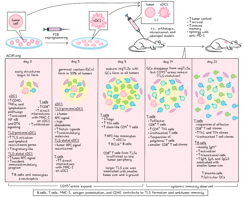
Tertiary lymphoid structures (TLSs) are associated with improved antitumor immune responses and immunotherapy efficacy. What triggers TLS formation and their composition remains largely unknown, as these are challenging to induce in preclinical models. Ascic, Chatelain, et al. investigated whether cDC1-reprogrammed tumor cells can induce immunogenic TLSs in various cancer types. Their results were recently published in Immunity.
Researchers first tested whether cDC1 reprogramming produces lasting antitumor immunity. Based on their prior work, they transduced tumor cells with a lentiviral vector encoding a cDC1-specific gene regulatory network (PU.1, IRF8, BATF3; PIB) and implanted them mixed 1:1 with parental cells in several subcutaneous tumor models. PIB reprogramming improved tumor control and long-term survival across all models. Surviving mice that were rechallenged rejected or showed delayed growth of tumors, suggesting immune memory. In orthotopic models, reprogramming produced more complete responses than anti-PD-1 therapy, increased tumoral T and B cell infiltration, and promoted memory formation. PIB reprogramming synergized with anti-PD-1 therapy in subcutaneous colon cancer and orthotopic melanoma. Finally, in the intracranial immunosuppressive glioblastoma model SB28, PIB reprogramming induced complete responses in 50% of mice, and synergized with anti-PD-1. Additionally, mice survived longer after rechallenge in the contralateral hemisphere.
In YUMM1.7 melanoma tumors 9 days after inoculation, densely nucleated structures surrounded by stromal-like cells were observed, with large numbers of immune cells and expression of the GC marker BCL6. These imgTLSs showed high infiltration by T and B cells. Imaging across multiple cancer models showed that cDC1 reprogramming induced these imgTLSs in all models at variable frequencies. Mutational burden did not affect the size of imgTLSs, but larger imgTLSs correlated with smaller tumor sizes at day 9 and reduced tumor growth.
To track imgTLS formation, researchers imaged YUMM1.7 tumors over time. Early structures appeared by day 3, 33% of tumors had germinal centers (GCs) by day 5, and all had mature imgTLSs with GCs by day 9. Immune-cell areas expanded from days 3–9. By day 14, GCs had disappeared, but CD45+ areas remained, consistent with the known process of TLS involution. Lymphoid infiltration rose on day 3, alongside fewer neutrophils and more monocytes. By day 9, there was an increase in APC-like monocytes and cDC1s.
To assess the role of antigen presentation in imgTLS formation, the researchers PIB-transduced mouse embryonic fibroblasts. This induced immune aggregates in tumors, but at lower frequencies than reprogrammed tumor cells, suggesting that antigen presentation contributes to imgTLS induction. Intratumoral delivery of an adenoviral vector encoding PIB in established YUMM1.7 tumors also induced lymphoid aggregates and mature imgTLSs by day 9.
To investigate how cDC1-like cells interact with T cells to coordinate the formation of imgTLSs, a sortase A-based system that biotinylates cells interacting with sortase A-expressing cells was used. Early biotinylation of cells within lymphoid aggregates was observed by day 3, and increased by day 5. Biotin-labeling was stronger within the imgTLSs than in surrounding tissue.
To establish whether T cell and cDC1 interactions were required for the formation of these TLSs, T or B cells were depleted. B cell depletion limited GC formation, while T cell depletion affected TLS maturation, resulting in a reduced GC area. CD40 agonist treatment could restore the imgTLS frequency in T cell-depleted animals, but not in B cell-depleted animals. Disruption of MHC and CD40 signaling showed that MHC-I and CD40 signaling were required for imgTLS formation, suggesting an important role for antigen presentation.
The researchers profiled transduced cells proximal or distal to imgTLSs. After day 3, the expression of a tumor-APC signature decreased in proximal cells, but persisted in distal cells through day 5. On day 3, proximal cells upregulated TLS initiation and lymphoid recruitment genes, while distal cells showed enrichment of genes related to antigen presentation. At day 5, proximal cells had high chemokine expression and increased Notch ligands and co-stimulatory molecules. Proximal cells acquired a migratory cDC1-like state at day 3, whereas distal cells adopted a resident immunostimulatory program. By days 3 and 4, both subsets rapidly induced lymphotoxin, TNFα, and CD40 pathways, and activated NF-κB and interferon signaling. These results suggested a spatially organized development program.
Compared with spontaneous immune aggregates in control tumors, imgTLSs contained fewer Tregs and more Tfh and stem-like CD4+ T cells. CD8+ T cells were enriched in imgTLSs on day 3, trafficking to peripheral tumor regions by day 9. BCL6-expressing B cells accumulated in imgTLSs.
To assess imgTLS-mediated systemic T cell responses, T cell states and clonotypes were profiled by TCR- and scRNAseq in imgTLS-containing and abscopal tumors, blood, and tumor-draining lymph nodes (tdLNs) on days 14 and 21. ImgTLS-containing tumors showed increased CD8+ effector and CD4+ Th1 populations and fewer exhausted cells. Abscopal tumors showed similar effects. At day 14, tumors with imgTLSs had polyclonal CD8+ T cell expansion. TCR clonotypes overlapped more between primary and abscopal tumors than with blood or tdLNs, suggesting migration between tumor sites. Compared with PIB-treated tumors, controls had larger CD8+ clones with similar phenotypes by day 14. By day 21, however, PIB expanded effector-state CD8+ clones, while control clones became exhausted. CD4+ Th1 cells followed similar trends, with exhaustion in controls. In PIB-treated tumors, Tfh and Th1 clonotypes were shared among primary and abscopal tumors and tdLNs.
At day 21, B cells increased 4.7-fold in PIB-treated YUMM1.7 tumors compared to control tumors, mostly retaining an IgM+ phenotype. Activated B cells, follicular DCs, and plasma cells also increased. Transitional B cells accumulated in tumors, while tdLNs had more activated B cells. Activated and cycling GC B cells increased in the blood, and plasma cells accumulated in abscopal tumors. IgM, IgA, and IgG3 concentrations increased and correlated with smaller tumors. Depleting B and plasma cells impaired tumor control in 36% of mice, which could partially be overcome by infusing day-28 plasma containing antibodies. These findings suggest that B cells promote antitumor effects through antibody-dependent and -independent mechanisms.
The data from this study show that reprogramming tumor cells to cDC1s can promote the formation of imgTLSs, thereby improving antitumor immunity. These models can be used to further optimize immunotherapies, particularly for immune-cold tumors.
Write-up by Maartje Wouters, image by Lauren Hitchings
Meet the researcher
This week, first co-authors Ervin Ascic and Camille Chatelain answered our questions.

What was the most surprising finding of this study for you?
EA: Clinical data had shown already that type 1 conventional dendritic cells are associated with improved survival across many solid tumor types, giving us confidence that this therapeutic approach could have broad, tumor-agnostic potential. However, evidence in brain tumors is very limited, and only recent studies have suggested that, even within the immune-privileged environment of the brain, dendritic cells may be linked to improved therapeutic responses. Therefore, the most surprising finding for me was the efficacy observed in the glioblastoma model. This result strengthened my confidence that this approach may help reshape how we understand and treat brain tumors.
CC: For me the most surprising finding was witnessing how profoundly the microenvironment shaped tumor-to-dendritic cell reprogramming. Although we introduced the same transcription factors, the reprogrammed dendritic cells acquired distinct states depending on their intratumoral positioning. It was even more exciting to find corresponding states and spatial patterns among endogenous dendritic cells in human tumor-associated tertiary lymphoid structures: DC-LAMP⁺ CCR7⁺ migratory dendritic cells were close to T and B cell niches, whereas CLEC9A-expressing dendritic cells were located further away. This really highlighted the relevance of our model for investigating tertiary lymphoid structures biology. Honestly, my cheeks were burning from how much I was smiling when we saw these results!
What is the outlook?
The next major step is to translate this approach from bench to bedside. This will require defining its limitations and potential mechanisms of resistance, identifying which patients are most likely to benefit, and establishing biomarkers to guide clinical use. Determining the precise molecular requirements through which dendritic cells initiate and maintain tertiary lymphoid structures would enable the modulation of tertiary lymphoid structures for distinct functional purposes, either tailored to specific tumor microenvironments and potentially to other diseases. The broader opportunity is to move beyond using tertiary lymphoid structures as a biomarker of treatment response to using them as a programmable tool to improve therapeutic outcomes.
If you could go back in time and give your early-career self one piece of advice for navigating a scientific career, what would it be?
EA: I would tell my early-career self not to worry too much about asking questions and starting discussions with more experienced researchers or peers. It is entirely acceptable not to know everything or to ask questions that may seem simple to step outside the comfort zone. Engaging with different perspectives and learning from more experienced researchers helps a lot to develop ideas or hypotheses and strengthen one’s confidence. I believe one needs healthy confidence and curiosity in science. Being open-minded and willing to ask for help and advice is an important part of that process.
CC: I would tell my younger self not to wait for the perfect plan, but simply to give things a try. Not everything will work out as expected, but every experience, including failure, is an opportunity to learn either about science or about yourself. So, be patient, stay curious, and dare to be a little bold. Learn to recognize your strengths and weaknesses, and don't hesitate to collaborate with others with skills and perspectives that complement your own. You don't have to do it all alone.


