

The ACIR team attended the SITC 2026 Annual Meeting in Phoenix, Arizona. This week’s extensive special feature covers select talks from the conference. We have organized the content by topics below.
Richard V. Smalley Memorial Award
Catherine J. Wu
William B. Coley Award Lecture
Eric Vivier
Keynote address
Ton Schumacher
Bispecifics
Harpreet Singh
John V. Heymach
Cytokines
Jeffrey A. Hubbell
Brett Pogostin
Immune checkpoint blockade
Lawrence Fong
Shailender Bhatia and Michael C. Lowe
CAR T cells
Marcela V. Maus
Mohammad Rashidian
Zachary Roberts
Kevin Reynolds
Cancer vaccines
Ryan Sullivan
Maha Ayyoub
William A. Freed-Pastor
Tumor immune microenvironment
Mikala Egeblad
Marco Colonna
Neil Chada
Metabolism
Greg M. Delgoffe
Richard V. Smalley Memorial Award
Getting Off the Rollercoaster: Personalized Cancer Vaccines Come of Age - Catherine J. Wu, MD – Dana-Farber Cancer Institute
After being awarded the Richard V. Smalley Memorial Award, Cathy Wu discussed the history and potential of personalized, neoantigen-targeted vaccines in overcoming tumor heterogeneity, which drives tumor evolution, resistance, and immune evasion, and is a major barrier to immunotherapy. While Wu described the pursuit of this strategy as a “rollercoaster” involving decades of research, the current wave of phase 3 clinical trials – enabled in large part by modern next-gen sequencing technologies, improved epitope prediction models, and advances with immune checkpoint blockades – is finally showing more reliable clinical benefits. In 2017, Wu and colleagues published their first early results from a phase 1 clinical trial establishing that personalized vaccines targeting up to 20 neoantigens using synthetic long peptides and supported by adjuvant poly-ICLC could mobilize vaccine-specific CD4+ and CD8+ T cell responses and induce epitope spread and long-term immune memory in melanoma. Similar results were published back-to-back by Ugur Sahin’s group using intranodally delivered mRNA encoding peptides, and shortly after, similar effects were demonstrated in glioblastoma (GBM). Since then, Wu and her team contributed to advancements in epitope prediction, disease monitoring, identification of immune markers of prognosis, evaluation of mechanisms of resistance following immunoselection, and understanding tumor-intrinsic and -extrinsic antigen landscapes. These advancements have opened up new avenues for treating more indications (including RCC and ovarian cancer) with better clinical outcomes. In current ongoing work, Wu and colleagues are investigating the impact of the timing of anti-PD-1 in responses to NeoVax in GBM. In a 4-arm study, 39 patients are being treated with pembrolizumab, starting either after vaccination (cohort 1), before vaccination (cohort 2), during radiotherapy, then a break, and again after vaccination (cohort 3). A fourth cohort (1B) with MGMT-methylated tumors is being treated on the same schedule as cohort 1, but with the addition of temozolomide (TMZ). Compared to matched historical controls, cohort 1B appeared to benefit more than cohort 1-3 (comprising patients with MGMT-unmethylated tumors), and cohort 2 (anti-PD-1 treatment started before vaccination) appeared to benefit more than cohorts 1 and 3. Ex vivo responses to vaccine peptides were associated with longer overall survival. Vaccine-reactive clonotypes were found to expand and persist up to two years. Cohorts 2 and 1B also showed strong increases in activating cytokines, chemokines, and chemoattractants upon vaccination. Analysis of paired tumor biopsies showed reduced Tregs in cohort 1B, possibly due to TMZ treatment. Further, vaccine-induced T cells were prominent in perinecrotic zones, suggestive of effective cytotoxicity. Looking at bulk transcriptional subtypes, the team saw associations between the mesenchymal subtype and response to anti-PD-1 monotherapy and survival, but these differences were largely leveled by vaccination. These continuing studies highlight how research in personalized vaccines can go from bench to bedside and back again to improve both understanding and outcomes. As numerous large, later-phase clinical trials (supported by the positive phase 2 and 3 trial results from Moderna and Merck) move forward into new clinical indications and approaches, advances in immunology research and technology will continue to serve as “engines of discovery” that propel immunotherapy forward.
Reading suggestion:
Ovarian cancer care gets personal in a phase 1 vaccine trial
William B. Coley Award Lecture
Harnessing NK Cells in Cancer Therapies - Eric Vivier, DVM, PhD – Marseille Medical School and Centre d’Immunologie de Marseille-Luminy
This year’s recipient of the prestigious CRI William B. Coley Award was Eric Vivier. As a leading scientist in the field of innate immunity, he focused his lecture on the biology of NK cells, their dysfunction, and novel therapeutic strategies using NK cell engagers. Juxtaposition of innate lymphoid cells (ILC1, 2, 3, and NK cells) and “adaptive lymphocytes” (CD4+ TH1, 2, 17, 22, and CD8+ CTLs) shows that the organization of the immune response is mimicked in these two systems. NK cells comprise prominent subsets: cytotoxic NK1 cells and cytokine-producing NK2 cells that are conserved across organs and species (mice and humans). While NK1 cells derive from canonical NK cell precursors, NK2 cells share lineage origins with ILCs. Vivier and colleagues recently defined 10 hallmarks of NK cell tumor immunity. A balance in the engagement of activating or inhibitory receptors on the cell surface of NK cells allows them to recognize “stressed” cells. NK cells do not require antigen-specific priming, can directly kill tumor cells, and have an excellent safety profile, as NK cells were selected in the species while T cells were selected in the individuals, leading to the possibility of GvHD. However, chronic stimulation within the tumor immune microenvironment through loss of MHC-I (an inhibitory ligand) and overexpression of stress ligands (which bind activating receptors) on tumor cells can lead to NK cell dysfunction. This functional exhaustion of tumor-infiltrating NK1 cells is marked by a transcriptional shift from the NK1 signature to the NK2 signature in mice and humans. While others study the treatment of cancer with ex vivo-conditioned, genetically manipulated, or CAR NK cells, Vivier and his team focus on harnessing endogenous NK cells in the fight against cancer. Similar to immune checkpoint blockade for T cells, the monoclonal antibody monalizumab can unleash NK cells by blocking the inhibitory interaction between HLA-E on tumor cells and NKG2A on NK cells. Monalizumab delivered positive results in phase II trials, and is currently being evaluated in a phase III study. In another approach to bypass immunosuppression of endogenous NK cells, Vivier and his team have developed multi-specific ANtibody-based NK cell Engager Therapeutics, ANKET. These tri- and tetra-specific NK cell engagers are designed to bind to NKp46 (a highly selective and very potent activating receptor on NK cells), CD16 (another activating receptor on NK cells), and a tumor-associated antigen to effectively redirect NK cells to cancer cells. Trispecific NK cell engagers targeting CD123 in acute myeloid leukemia and BCMA in multiple myeloma are currently in clinical development. In addition, a tetraspecific NK cell engager targeting CD20 and incorporating an IL-2v domain lacking CD25/IL-2Rα binding is being studied in clinical trials. Next, Vivier presented preclinical data for tetraspecific IFNα2-armed ANKETs. IFNα2-armed ANKETs outperformed IL-2v-armed ANKETs in vitro and in vivo in terms of safety and efficacy. In contrast to IL-2v-armed ANKETs, IFNα2-armed ANKETs do not stimulate NK cell proliferation, but induce robust local NK cell activation, driving superior tumor control in a Raji lymphoma model. The IFNα2 moiety of the ANKETs has a strong effect on NK cells, but since the IFNα2-receptor IFNAR1/2 is also expressed on tumor cells and it is known that binding of IFNα to FNAR1/2 on tumor cells can induce apoptosis and immunogenic cell death, the IFNα2 moiety of the ANKETs could also function as an antibody–drug conjugate. Linking IFNα2 to the tumor antigen binder re-adresses the cytokine within the ANKET to tumor cells, leading to tumor cell lysis in vitro and in vivo. Finally, Viver quickly touched on ILCs, which are mainly tissue-resident. Together with other tissue-resident immune cells, these cells may define the spatial organization of immune responses within specific tissues – “regional immunity” – which may also have an impact on normal organ physiology. Many assays used to evaluate drug candidates may not accurately reflect the phenotype and fitness of tissue-adapted immune cells, and toxicities of interventions may reflect disruptions of the regional immunity. In short, Vivier underscored the expanding clinical potential of harnessing NK cells in cancer immunotherapy. And FYI, next to the famous band “The Checkpoints”, another band has entered the stage: “The Engagers”, with Eric Vivier on the electric guitar.
Reading suggestion:
A tetraspecific NK cell engager armed with IL-2v targets B-NHL
Keynote Address
T Cell Recognition of Human Cancer: “Cracking the Code” - Ton Schumacher, PhD – Netherlands Cancer Institute
In his keynote lecture, Ton Schumacher focused on “cracking the code” – the challenging goal of attempting to predict TCR specificity based on the sequence of the TCR only. If successful, this would have enormous benefit not only in cancer and autoimmunity, but potentially in any other disease where T cells are involved. Ultimately, successful immunity depends on the interaction of a distinct set of TCRs with a distinct set of antigens presented by abnormal cells, leading to their specific killing. Furthermore, the interacting partners, TCR and antigen, are unique to each patient. Being able to very easily determine the identities of each of those players is the key problem to solve. Schumacher believes he and his team have made progress toward that goal, and described two technologies supporting that belief. Furthermore, he showed how such approaches could explore an immunological question regarding immune-inflamed vs. immune-excluded tumors. The mathematical magnitude of TCR::peptide:MHC (TCR:pMHC) pairs is huge, and has been approached by others using sequence-based algorithms or structure-based modeling (using Alpha-fold 3 [AF3]), but with only modest results. Asking whether this was a technology or problem with the underlying data – much of which came from the VDJdb database, which collates TCRs with specificity to known antigens – Schumacher’s team developed and applied a high-throughput robotics-assisted TCR synthesis approach (T-RAP) and synthesized ~4,000 TCRs reported in the VDJdb database to interact with a given peptide:MHC. Engineering those TCRs into reporter cells as a library, and using that library to probe the reported individual peptide:MHC reactivity directly addressed the assignment quality. Overall, only about 50% of the annotated TCR:pMHC pairs validated, which may explain at least part of the problem of approaches to data. The newly generated, higher quality data subset demonstrated improved predictivity in properly pairing the tested epitope/TCRs, but also TCR:pMHC complexes with no prior structural information or for novel neoantigens, eliminating the possibility of data leakage. Training AF3 with the improved database, employing both consensus and iterative approaches (sequence clustering and structural docking modeling), generated a new algorithm, TCR-dock3, which further improved predictivity looking at multiple external datasets, including even murine TCR:pMHC and in silico-designed TCRs. To apply these new technologies to an immunological problem, Schumacher addressed the question of the distinction between T cells that extravasate the blood, but accumulate at the tumor rim in “excluded” tumors (usually unresponsive to immune checkpoint blockade [ICB]), and those that infiltrate a “hot” tumor, (usually responsive to ICB). Cold tumors significantly outnumber hot tumors at baseline, so understanding and overcoming this problem could significantly improve ICB response. RNA and DNA sequencing of subsets of tumors based only on the hot vs. cold immunohistochemistry distinction revealed comparable tumor mutation burden and the expected dominance of TGFβ in cold tumors, or interferon signaling in hot tumors. Using T-RAP to prepare TCRs from each tissue sample, and the published HANSolo approach to screen those TCRs against a library of patient-specific neoantigens, tumor-associated antigens (TAA) and viral antigens (control) revealed that the repertoire of targeted neoantigens and TAAs was comparable between tumor types, so infiltration was not target-dependent. To address the quantitative difference and pair TCRs with antigens on a population level, the team developed PAIR-Scan to allow library-on-library (epitope and TCR) screening at scale. The procedure capitalizes on the enhanced pairing of reactive T cells with cognate APCs to generate doublets, a calcium recorder to select those pairs properly signaling, andRNA sequencing of the doublets to simultaneously identify epitope and reactive TCR, and to capture the transcriptomic state of those cells. Scanning 40,000 combinations of the 200 most highly expressed epitopes and the 200 most abundant TCRs derived from a patient with stage IV melanoma revealed the (expected) small subset of hits, most of which validated. Early results of applying PAIR-Scan to cold and hot tumors suggests that the number of reactive T cells is comparable in both inflamed and excluded tumors. Differences, however, appear in the inferred phenotypic states of these cells, particularly in the T cell expression of signaling components such as, chemokines and, unexpectedly, TGFβ, while cells in both tumor types appear to have comparable TCR signaling activity. These results point to the value of overcoming the exclusion of T cells in cold tumors to capitalize on the repertoire, number, and phenotypic state of the T cells sitting on the tumor rim.
Reading suggestion:
T-RAP – a new tool for predicting TCR-pMHC reactivity
Bispecifics
Targeting PRAME with IMA402, a Novel Bispecific T-Cell Engager (TCER) - Initial Clinical Results - Harpreet Singh, PhD – Immatics
Harpeet Singh discussed therapies targeting PRAME, which is an intracellular protein that can be targeted by TCRs recognizing a PRAME peptide presented through HLA*A2:01. PRAME is homogeneously expressed in the tumor tissue across over 50 solid tumor types, and has been associated with poor survival. Immatics has developed three therapeutic products targeting PRAME: two autologous cellular therapies and one T cell engager using a high-affinity, highly specific TCR that recognizes PRAME. The autologous cell therapy, anzu-cel, was tested in a phase 1b trial in heavily pre-treated patients with metastatic melanoma, and showed an overall response rate (ORR) of 56%, with a median duration of response of >18 months. The other cellular therapy is IMA203CD8, which has shown broad activity in a dose-escalation phase 1a trial across several cancer types, including melanoma, synovial sarcoma, and gynecological cancers. The ORR was 41%, and 55% in the group with the right target dose. Toxicity for both cellular therapies was manageable and comparable to other cellular therapies. The T cell engager IMA402 is a bispecific with three components. It includes a TCR domain derived from the same parental TCR as the cell therapies. The other specificity is an antibody fragment that recognizes the CD3/TCR complex; this is deliberately low-affinity to allow for higher doses of IMA402, as the bispecific first has to bind the tumor cells before it can bind to T cells, preventing non-specific T cell activation in the periphery. The third component is an antibody-like molecule that extends the half-life of IMA403 to over one week. In a Phase 1a dose-escalation trial (n=126), IMA402 monotherapy or IMA402 plus pembrolizumab was assessed in heavily pre-treated patients (including immune checkpoint blockade [ICB]) with advanced solid PRAME+ tumors and high tumor burden. Toxicity was manageable, with some cases of transient lymphopenia and low-grade cytokine release syndrome (CRS). The ORR was 25%, with a disease control rate of 63%. PRAME expression was a biomarker for response, and all confirmed responders had high PRAME expression. Across multiple cancer types, the PRAME-high group had an ORR of 32%. At a median follow-up of >17 months, the median duration of response was >12 months. Patients dosed at the recommended phase 2 dose had a 1-year survival rate of 91%, although this data is still premature, with a median follow up of 8.8 months. A liquid biopsy method has been developed to reliably detect PRAME in the blood as a companion diagnostic. Further, Singh noted the development of another T cell engager targeting MAGEA4/8 (IMA401). In a recently started phase 1a trial, the combination of the PRAME and MAGE engagers is being assessed in patients with squamous non-small cell lung cancer, as 90% of tumors express either of these targets, and over 60% express both, so IMA401 and IMA402 may have synergistic effects. Further, combinations with ICB and the chemotherapeutic decitabine are being assessed, the latter because it induced PRAME upregulation in preclinical data.
Reading suggestion:
MAGE-A4/MAGE-A8-targeted TCR-based bispecific T cell engager in recurrent and/or refractory solid tumors: a phase 1 trial
Biology and Clinical Results of Anti-VEGF + PD(L)-1 Bispecifics - John V. Heymach, MD, PhD – MD Anderson Cancer Center
VEGF/PD-(L)1 bispecific therapeutics are advancing rapidly across multiple phase 2 and 3 clinical trials for various solid cancers. John Heymach outlined that these bispecifics have been engineered in various antibody-based formats, but all feature non-competitive bivalent binding to both VEGF and PD-1 or PD-L1, with a silent Fc region. Heymach then focused on the rationale for simultaneous engagement of both targets by a singular bispecific. VEGF blockade alone helps counteract tumor-driven immune suppression by affecting M2 macrophage polarization, MDSCs, and DC maturation, mitigating T cell exhaustion, and normalizing vascular tortuosity (turns and bends of the blood vessels) and permeability to induce tumor necrosis. This immune modulation occurs primarily through VEGF receptor 1 rather than VEGF-R2, which is predominantly found on vascular and tumor cells. VEGF/PD-1 bispecifics, such as ivonescimab, promote cooperative binding to PD-1, driven by VEGF-induced oligomerization (“daisy-chaining”). This structural oligomerization markedly slows dissociation rates, enhances PD-1 internalization, and significantly boosts IFNγ secretion in the presence of soluble VEGF. Similarly, soluble PD-1 enhances ivonescimab-induced VEGF-R2 signaling blockade. Cooperative binding and enhanced functionality was also observed with the VEGF/PD-1 bispecific JS207 and the VEGF/PD-L1 bispecific pumitamig. The randomized phase 3 HARMONi-2 clinical trial, conducted in China, evaluated ivonescimab versus pembrolizumab as a first-line treatment for patients with PD-L1-positive (≥1%) NSCLC. At median follow-up of 8.67 months, ivonescimab nearly doubled the progression-free survival (PFS) compared to pembrolizumab (11.1 months vs. 5.8 months; HR 0.51) alongside a significant overall survival (OS) benefit (HR 0.73). The pembrolizumab-only arm appeared to perform similarly to past studies in this patient population, and ivonescimab showed a trend toward greater activity in patients with lower PD-L1 levels. While grade ≥3 treatment-related adverse events (TRAEs) were almost twice as high for the ivonescimab-arm, they were manageable and did not lead to increased discontinuation or death. Interestingly, TRAEs such as proteinuria and hypertension occurred at lower-than-expected rates compared to traditional bevacizumab. In the randomized phase 3 HARMONi clinical trial, ivonescimab plus chemotherapy demonstrated a strong PFS benefit (HR 0.52) compared with placebo plus chemotherapy in EGFR-mutant NSCLC post-TKI therapy. Consistent efficacy was also observed in the randomized phase 3 HARMONi-6 clinical trial, where ivonescimab plus chemotherapy outperformed anti-PD-1 tislelizumab plus chemotherapy in first-line squamous NSCLC (PFS HR 0.60 and OS HR 0.66). Beyond lung cancer, the HARMONi-GI-1 trial in patients with advanced biliary tract cancer demonstrated that ivonescimab plus chemotherapy significantly improved OS (HR 0.75) compared to durvalumab plus chemotherapy. And finally, in two studies led by Heymach in patients with extensive-stage SCLC, pumitamig with or without chemotherapy achieved response rates of 85%. Heymach concluded that preclinical and clinical data strongly support that VEGF/PD-(L)1 bispecifics provide synergistic effects beyond separate or monotherapy blockades, with ongoing clinical trials like HARMONi-3 and -7 poised to further establish their clinical potential.
Reading suggestion:
Elimination of large tumors in mice by mRNA-encoded bispecific antibodies
Cytokines
Conditional Deposition of Cytokines and Chemokines in the TME via Enzymatic Reactivity - Jeffrey A. Hubbell, PhD – New York University
Rather than using enzymes in the tumor microenvironment to demask cytokines and break bonds, Jeffrey Hubbell and his team used a novel approach of leveraging enzymes naturally upregulated and activated in solid tumors to form covalent bonds and deposit cytokines in the TME. The collagen or elastin cross-linking enzymes lysyl oxidase (LOX) and LOX-like 1, 2, 3, and 4 are upregulated under hypoxic conditions in solid tumors, depend on elevated levels of copper (the copper transporter ATP7A is upregulated in solid tumors), and are further activated by BMP1 (also upregulated in solid tumors). To deposit a therapeutic agent such as a cytokine selectively in the tumor upon intravenous or distant subcutaneous administration, Hubbell and colleagues engineered cytokine fusion proteins containing the 7S domain of type 4 collagen, a substrate of LOX and LOX-like 1-4. Subcutaneous injection of LOX-targeted IL-7 and IL-12 (LOD–IL-7 and LOD–IL-12) fusion proteins on the contralateral site of the tumor, together with anti-PD-1 treatment, demonstrated a significant survival benefit in s.c. MC38 and B16F10 mouse models. Immune phenotyping of B16F10-bearing mice treated with LOD–IL-7 and LOD–IL-12 (without anti-PD-1) revealed elevated CD8+ T cell numbers, enhanced functional CTL markers (including TCF1 as a marker of stemness), decreased markers of exhaustion, and an increased ratio of CD8+ T cells to Tregs. Compared with wild-type IL-7 and IL-12, the targeted cytokines yielded sustained, increased levels of intratumoral IFNγ. In an autochthonous, less inflamed melanoma model, although with adequate LOX activity, the distant s.c. injection of LOD–IL-7 and LOD–IL-12 with anti-PD-1 led to improved progression-free survival. Next, Hubbell showed that distant s.c. LOD–IL-7 and LOD–IL-12 injection in an orthotopic renal cell carcinoma model or i.v. injection of the targeted cytokines plus anti-PD-1 in an immune-exhausted orthotopic hepatocellular carcinoma model can affect these “central” tumors and increase long-term survival while preserving normal renal and hepatic function, respectively. Finally, Hubbell was able to demonstrate that this approach would even work for chemokines. Despite distant s.c. administration, LOD–CCL5 plus anti-PD-1 successfully induced chemotaxis and infiltration of cytotoxic CD8+ T cells in an MC38 tumor model due to selective deposition in the TME, and led to complete responses in 2 of 6 animals.
Reading suggestion:
Targeting tumor collagen hits the immunotherapeutic sweet spot
CD45-Targeted IL-12 Reveals a cDC2–CD4⁺ T Cell Axis as a Driver of Lung Tumor Immunity - Brett Pogostin, PhD – MIT
Non-small cell lung cancer has few effective therapies, and although immune checkpoint blockade (ICB) does provide tumor control and a survival benefit, the effect is modest, and more effective therapies are needed. Brett Pogostin built on prior work from the Spranger lab showing that defective IL-12 signaling in tumor-draining (mediastinal) lymph nodes (mTDLN) resulted in dysfunctional T cell priming, and hypothesized that targeting IL-12 to immune cell-rich lymph nodes with an IL-12 molecule linked to an anti-CD45 antibody (αCD45:IL-12) might overcome the high systemic toxicity of IL-12. Additionally, molecules tethered to surface CD45 showed reduced receptor-mediated internalization, potentially extending cytokine signaling. In the KP.SIY GEMM model (both as autochthonous and transplantable), multiple intraperitoneal (i.p.) doses of the αCD45:IL-12 immunocytokine resulted in improved and durable survival. Mechanistically, there was significant accumulation of the drug in the mTDLN and at day 21 in the lungs of long-term survivors. Interestingly, this therapy was effective despite the absence of MHC-I expression on these tumor cells. Treatment improved tumor-specific T cell numbers, which were almost entirely CD4+ T cells. Depletion of CD8+ T cells after therapy had no effect on tumor control, while depletion of CD4+ T cells completely abrogated the effect. CD4+ T cells can be primed by either cDC1s and cDC2. Treatment of mice lacking cDC1s still resulted in T cell priming and tumor control, while mice lacking cDC2s were defective in both functions, pointing to a critical role for cDC2s in this therapeutic effect. Surprisingly, despite knockout of MHC-II in cDC2s, the cells were still able to prime T cells, indicating that there was another source of MHC-II, perhaps tumor cells themselves. Knockout of MHC-II in both tumor cells and cDC2s demonstrated loss of priming, suggesting that cDC2s were acquiring MHC -II complexes from tumor cells via cross-dressing. Acquisition of MHC-II from tumor cells by cDC2s was directly observed using flow cytometry. Finally, looking at the distribution of MHC class I and II expression on human NSCLC tumors, about 33% of patients expressed normal levels of MHC-II and low levels of MHC-I, a substantial sub-population which might not be expected to respond to αPD-1/L1 therapy, but might respond to αCD45:IL-12.
Reading suggestion:
CCR7+ activated dendritic cells are essential for spontaneous and immunotherapy-driven anti-tumor immunity
Immune checkpoint blockade
Distinct Immune Responses Induced by Different Forms of Radiotherapy Combined with Immune Checkpoint Inhibition - Lawrence Fong, MD – Fred Hutchinson Cancer Center
Lawrence Fong discussed recent efforts to sensitize ICB-refractory prostate cancer with the addition of various forms of radiotherapy, including systemic 223Radium, systemic PSMA-targeting 177Lu, or external beam radiotherapy (EBRT). 223Radium, which is approved for use in metastatic castration-resistant prostate cancer (mCRPC) with bone metastases, is known to improve overall survival and delay skeletal events. In a recent clinical trial in combination with pembrolizumab (anti-PD-1), the combination showed limited differences compared to pembrolizumab alone, with similar PFS and OS. Further, CD4+ and CD8+ T cell infiltration were similar, and 223Ra did not contribute to any increase or decrease in immune markers in circulating T cells. However, immune markers did differ by response status, with responders showing lower circulating Ki67+ T and myeloid cells at baseline, and higher circulating Tim3+ T and myeloid cells by week 9. In another trial, 177Lu-PSMA617 (Pluvicto; always only a single dose) was evaluated in combination with anti-PD-1 (always multiple doses 3 weeks apart) on various treatment schedules. Patients were treated with either Pluvicto alone, both treatments starting at the same time, or anti-PD-1 starting prior to a single dose of Pluvicto. The objective response rate was high across treatment groups, with some patients experiencing long-term responses. Unlike 223Ra in the prior study, Pluvicto showed immunogenicity as a single agent, and in combination, it enhanced the immunogenicity of anti-PD-1, with differential responses at the single-cell level. In responders, effector CD8+ T cells, γδ T cells, and NK cells increased in frequency and became more activated. Finally, Fong discussed a trial evaluating EBRT in combination with anti-PD-1 and standard-of-care hormonal therapy. Single-cell analysis of pre- and post-treatment biopsies showed that combination treatment reduced CD8+ T cells and increased myeloid cells. Meanwhile, intratumoral T cells shifted from effector to naive phenotypes. Clinical responders had increased intratumoral T cell clonality, and responses were associated with both induced and persisting tumor-homing cells. These results highlight several potential benefits and drawbacks of combining different forms of radiotherapy with anti-PD-1.
Reading suggestion:
Turning up the heat: iSBRT and immunotherapy induce immune reprogramming in cold breast tumors in the clinic
Radiotherapy from the inside out? TRT enhances immunotherapy
Debate: Pros and Cons on Time of Day and Checkpoint Inhibitors - Michael C. Lowe, MD, MA – Emory University School of Medicine and Shailender Bhatia, MD – University of Washington/Fred Hutch
An amusing debate was organized between Michael Lowe and Shailender Bhatia on whether the time of day matters for immune checkpoint blockade (ICB) infusion. Lowe argued for the “pro” side, that the timing of immunotherapy is clinically important. His arguments included various preclinical and retrospective data suggesting a correlation between early infusion times and better outcomes. Since it is a non-toxic, well-tolerated intervention, Lowe argued that patients deserve to have access to it if it provides any clinical benefit. Lowe noted that he was aware that Bhatia had found in a retrospective study that there was no PFS or OS difference in their clinic regarding early or later infusion times. Lowe argued that this was only one study with a limited number of patients (n=216), in contrast to a slightly larger study (n=299) led by Zach Buchwald at Emory, which showed an impact with a hazard ratio (HR) of 2.16. He argued that any other intervention would immediately garner much attention with such a HR. Two large meta-analyses with over 6,000 patients in total showed improved overall survival with early time of infusion (HR 0.5 and 0.61, respectively). In preclinical models, time of day of infusion was also associated with improved outcomes, with some infusion times being equal to controls. Further, time of day correlated with CD4+ and CD8+ T cell phenotypes found in human melanoma tissue. To take wind out of Bhatia’s sails, Lowe argued providently that previous meta-analyses and post-hoc studies failing to show a timing impact did not investigate whether time of day matters in an a priori fashion. According to Lowe, the recent traction of the LungTIME-C01 trial publication from Nature Medicine serves as a call to continue rigorous scientific investigation, rather than abandoning the hypothesis. Prospective trials are actively enrolling patients across multiple tumor types globally to properly test the impact of the time-of-infusion intervention. Bhatia, taking on the “con” side, confirmed that the large retrospective studies showing benefit were important data, and that there is evidence of immune cells in tumors fluctuating over different times of the day. However, he noted that this is looking at a very narrow part of the immune system, while circadian biology is complex, with various factors fluctuating throughout the day. Therefore, the whole body and immune system should be considered when discussing interventions. The pharmacological and dynamic profiles of ICB therapy raise significant doubts regarding whether the timing of administration meaningfully affects therapeutic outcomes. With nivolumab (anti-PD-1), the antibody is present in the serum at minimum effective concentrations for as long as 8 or 9 months after an infusion. Therefore, Bhatia asked if anti-PD-1 is present for so long, does the timing in the first hours really matter, and if so, why are patients put through repeated infusions for years? Further, he argued that it is not an easy intervention for clinics. In his center, infusions happen all day long, and there would be limited availability to offer patients if infusions could only be performed in the morning. He also noted that other disparities in data may affect efficacy as well. For instance, the NCI/SOWG trial S1404 found that patients living farther from a treatment center are known to have worse outcomes and are more likely to receive afternoon treatments. Therefore, Bhatia agreed that prospective randomized controlled trials are needed, given the heterogeneity in the current data in terms of treatment regimens, timing cutoffs, and tumor types, and a publication bias towards positive results. However, he argued that the current prospective studies will not answer all questions, given their intended treatment-combination regimens. Bhatia reviewed a 10-year retrospective analysis from his center only including patients with front-line ICB and no chemotherapy, focusing on immediate effects like tumor shrinkage, and observed no correlation with time of infusion. He concluded with the satirical observation that tumor response rate correlated more strongly with the day of the week, specifically Mondays, because immune cells recharge over the weekend and are not weakened.
Reading suggestion:
TMEs show night and day differences in accordance with circadian rhythms
Working 9 to 5: circadian rhythm in immune cells affects immunotherapy
CAR T cells
Learnings From a CAR-T Driver Into Solid Tumor - Marcela V. Maus, MD, PhD – Massachusetts General Hospital
Marcela Maus presented efforts aimed at improving efficacy of CAR T cell therapy in solid tumors. Her early efforts with an EGFRv3-targeted CAR-T in glioblastoma were limited by heterogeneity in tumor antigen expression and infiltration of Tregs. To overcome these challenges, a design platform was developed in which instead of modifying the T cell with just the CAR, additional transgenes were added, such as a transgene for a T cell-engaging antibody molecule (TEAM). The idea behind this was to leverage the T cell to reach inaccessible tumors and manufacture the TEAM locally, reducing the risk of on-target, off-tumor toxicity and enabling targeting of more than just one tumor antigen. TEAM can also then redirect Tregs to degranulate to and kill tumor cells. The strategy was tested in a clinical trial with a vector construct encoding the transgenes for the EGFRv3-directed CAR with a 4-1BB signaling domain, and a T cell engager with a scFv targeting wild-type EGFR and a low-affinity scFv targeting CD3, leveraging passenger T cells to contribute to the antitumor response. The first 3 patients with recurring glioblastoma were treated with CARv3-TEAM-E, and they experienced rapid tumor regression. However, two patients experienced recurrences, and received a second dose. This was ineffective, and the CAR-T did not engraft in the cerebrospinal fluid (CSF). Furthermore, in all patients, the CARv3-TEAM-E cells were cleared from the CSF within ~6 weeks. Maus and team hypothesized that increased persistence was needed for sustained responses, and that a potential immune response against the CAR and/or TEAM molecules developed after the injection of the cell therapy into the CSF without prior lymphodepletion. Indeed, all patients had developed anti-CAR and, in 2 of 3 cases, anti-TEAM antibodies. Instead of changing the vector, Maus and her team decided to treat the patients with lymphodepleting chemotherapy 3 days before each infusion. Patients were re-dosed on schedule every 4-6 weeks for up to 6 doses with CARv3-TEAM-E. While benchmark data suggest a median overall survival (OS) at first recurrence of 8 months, in this study the 12-month OS was 80%, with the median OS not yet reached, though the study sample size was small (n=10). Lymphodepletion reduced anti-CAR/anti-TEAM antibody formation, enabling the re-dosing and re-engraftment. Maus also found that, while the CNS is considered an immune-privileged site, even intra-CNS injections could prime immune rejection of the minimal foreign drug product. With these encouraging survival findings, Maus and colleagues are now investigating the addition of rituximab to the conditioning regimen to further reduce anti-CAR/anti-TEAM antibody formation. A second approach Maus discussed was assessment of the best gene knockouts in CAR-T to increase their persistence. For this, the MARIO-CART platform was developed, which includes an sgRNA library targeting genes that have been shown to confer some improvement to T cell function when knocked out. This was assessed with a BCMA-targeting CAR in an NSG mouse model for multiple myeloma. The top gene knockout hits varied across time points, likely due to changes in selective pressure over time. Further, for solid tumors, the top genes differed from hematological cancers, separating even more over time. Maus argued that in patients, all CARs need to be tested at once to determine which is the best CAR for that patient, and that tracking individual genetic markers is feasible with this method. Due to the high number of potential innovations to CAR-T, it is challenging to develop strong clinical data and compare products. Therefore, she noted that a regulatory structure is needed to enable innovation in cell therapies and to more quickly identify the best innovations.
Reading suggestion:
Dimming IFN signaling to brighten CAR-T persistence and efficacy
CAR-Enhancers: Programmable Boosters for Durable Cell Therapy - Mohammad Rashidian, PhD – Dana-Farber Cancer Institute
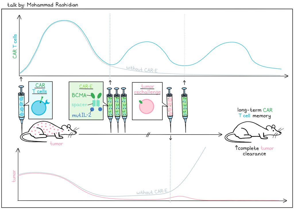
CAR T cell therapy can induce strong responses and frequently, tumor clearance for hematological cancers, but patients very often relapse with antigen-positive disease, as CAR T cells fail to persist long enough to clear elusive micrometastases. To enhance CAR T cell persistence and long-term functionality, Mohammad Rashidian and colleagues engineered a CAR enhancer (CAR-E) molecule consisting of the CAR’s target antigen (to ensure specific delivery to CAR T cells in circulation) bound to a low-affinity IL-2, with a spacer in between. This design had a short half-life in circulation, limiting its effects on normal T cells and enabling flexible dosing for safety. In a CAR T cell model using BCMA as the target antigen, administration of a BCMA–IL-2 CAR-E two weeks after CAR T cell administration induced robust re-expansion of CAR T cells and improved tumor control 3 days later. When mice were rechallenged at day 60 and then treated again with the CAR-E, similar re-expansion of the initial population of CAR T cells was observed, resulting in rejection of rechallenge. Investigating BCMA–IL-2 with different IL-2 variants showed that IL-2 signaling strength and its ability to induce STAT5 signaling was critical to the re-expansion and antitumor efficacy induced by CAR-E, with some molecules resulting in a 10,000-fold expansion. Rashidian also identified an optimized dosing schedule that sustained CAR T cells in circulation within a safe therapeutic window while preventing exhaustion. Mice that were rechallenged repeatedly continued clearing tumors, demonstrating that re-expanded CAR T cells maintained functionality long-term. Further analysis showed that a stem-like naive phenotype was maintained, demonstrating that T cells were of high quality, even in models using CAR T cells derived from patients with multiple myeloma. Looking at CAR T cell subsets, the researchers found that the CAR-E preferentially impacted CD8+ T cells, and transcriptomic data revealed a unique profile in CAR T cells after CAR-E administration that was distinct from profiles observed after exposure to antigen, low-affinity IL-2, a combination, or wild-type IL-2, suggestive of a totally distinct pathway. Upon further investigation, they found that the CAR-E molecule prolonged IL-2 signaling (despite its short half-life), remaining bound to the cell surface for hours rather than being rapidly internalized within minutes (as with unbound low-affinity or wild-type IL-2). Interestingly, signaling did not appear to be occurring through the IL-2 receptor, but rather through the CAR itself. Similar results were observed when IL-2 was swapped out for a mutated version of IL-21, which has no ability to expand T cells when bound to the endogenous IL-21 receptor. In vivo, the BCMA–IL-21 enhancer also induced robust CAR T cell expansion. While further investigation is needed to fully understand this mechanism, a clinical trial has been designed to test the potential of the BCMA–IL-2 CAR-E to enhance the long-term efficacy of existing CAR T cell therapy.
Reading suggestion:
A CAR enhancer increases the activity and persistence of CAR T cells
Allogeneic CAR T: A Platform Built for Patient Access – in Cancer and Beyond - Zachary Roberts – Allogene Therapeutics
Zachary Roberts discussed three allogeneic cell therapy programs that Allogene Therapeutics is developing to provide faster and more simplified access for patients than autologous products offer. He noted that only about 15% of patients who could benefit from a second-line autologous CAR-T therapy actually receive it due to access challenges. The first therapy aimed at overcoming some of these challenges is cema-cel, a CD19-targeting allogeneic CAR-T for large B-cell lymphoma (LBCL). This application is based on recent results indicating that the risk of relapse following first-line therapy is highly dependent on the minimal residual disease (MRD) state, which can be measured using a circulating tumor DNA (ctDNA) assay. The current standard of care for LBCL includes front-line induction chemotherapy regimens, with approximately 90% of patients achieving remission. This is followed by a “watch and wait” approach until relapse, which occurs in approximately a third of patients. In the randomized controlled ALPHA3 trial, rather than observation, patients received a ctDNA assessment of MRD at the end of front-line therapy, and those with detectable MRD were then treated with allogeneic off-the-shelf CAR-T. Due to low disease burden, toxicity was expected to be limited. Among 12 patients, 58.3% had MRD clearance after cema-cel, versus 16.7% in the control arm. Early safety data suggest limited toxicity, with no hospitalizations or need for steroid/tocilizumab treatment for toxicity. The second program is allo-316, a CD70-targeting allogeneic CAR-T assessed in metastatic, unresectable, relapsed clear cell renal cell carcinoma, which expresses high levels of CD70. CD70 is a dynamic marker of T cell activation and is upregulated during CAR-T manufacturing, risking fratricide by cells that target CD70-expressing cells (CD70-CAR-T cells). The researchers overcame this by identifying anti-CD70 Fabs, which bind membrane-distal epitopes on CD70, to construct the CAR. On an individual CAR T cell, the CD70-targeted CAR binds and blocks CD70 in cis, protecting it from fratricide by other CD70-targeted CAR-expressing cells. Targeting CD70 may also help prevent allo rejection, as recipients’ host T and B cells upregulate CD70 in response to the HLA-unmatched allo product, which are then cleared by the CAR-T. This has been shown to improve expansion and survival in vitro. In the TRAVERSE phase 1b trial in renal cell carcinoma with the ALLO-316 CAR targeting CD70, among patients with at least 50% CD70 expression, 44% achieved a minimum of 30% reduction of their target lesion, with an ORR of 30%, resulting in durable responses (18 months). Allo-316 showed excellent expansion and persistence compared to published results for autologous CARs. The final program is ALLO-329, a CD19- and CD70-targeting allogeneic CAR-T for autoimmune diseases. Mouse work showed that while the CD19 CAR-T required lymphodepletion for persistence, the CD19/CD70 CAR-T did not. Therefore, ALLO-329 is now being assessed in the RESOLUTION basket trial for several autoimmune disorders, in which it is being tested with low or no lymphodepletion states.
Reading suggestion:
Allogeneic CD19 CAR T cells armed with an anti-rejection CD70 CAR overcome antigen escape and evade alloimmune responses
Off-the-shelf allogeneic CAR-T cells one step closer to becoming a reality
Adaptive anti-CAR immunity compromises the durability of GD2-CAR T cell therapy in diffuse midline glioma - Kevin Reynolds – Stanford Cancer Institute
Diffuse midline glioma (DMG) is an aggressive pediatric cancer with poor survival. Given its high GD2 expression, GD2 CAR-T therapy has been developed for DMG. In a phase 1 study of first-line CAR-T treatment, patients received one cycle of lymphodepletion, followed by an intravenous infusion and then sequential intracerebroventricular injections of CAR-T cells. Patients had initial clinical benefit, with shrinking tumors, but ultimately progressed. Kevin Reynolds presented data on the potential treatment resistance mechanisms. Single-cell analysis of T and B cells in the cerebrospinal fluid (CSF) showed major clonal expansion of endogenous CAR-negative CD4+ and CD8+ T cells, with low persistence of CAR-positive cells. The landscape of plasmablast BCRs diversified, with various isotypes represented: the dominant isotypes were IgA1 and Ig1, with some presence of IgA2 and IgG2. This led to the finding that patients developed human anti-CAR antibodies (HACAs). Despite repeated infusions, CAR-positive cells decreased, while plasma HACAs increased. A novel ELISA assay with patient plasma and purified GD2 CAR protein confirmed the increased production of IgG and IgA1 HACAs over time. Patient outcomes were worse with more HACAs present. Expressing the highest-occurring plasmablast BCRs from three patients as monoclonal antibodies, 15 of 18 clones were found to specifically bind the GD2 CAR, and the class switch from IgA to IgG had no impact on binding. In a co-culture of CAR-T cells with GD2-expressing NALM6 cells and patient pools of HACAs, CAR T cell blockage and dose-dependent blocking of tumor killing was observed. Further research showed that HACA-mediated effects included both antibody-dependent cellular cytotoxicity (ADCC) and phagocytosis (ADCP). In vivo, HACAs also cleared CAR T cells in NSG mice engrafted with mock or CAR T cells. HACAs with a “dead” Fc domain, which cannot mediate cellular clearance, had no effect in this model. In sum, the data suggest that CAR immunogenicity is a mechanism of resistance, and efforts are now focused on modifying the therapy to prevent this effect.
Reading suggestion:
GD2-CAR T cell therapy shows promise against deadly pediatric cancer in early clinical trial
Cancer vaccines
Spatial profiling identifies response-associated tumor-immune niches and treatment-induced remodeling with mRNA-4359 plus pembrolizumab in melanoma - Ryan Sullivan, MD – Mass General Brigham Cancer Institute
Ryan Sullivan discussed early phase 1/2 clinical trial results for patients with metastatic melanoma who were treated with mRNA-4359 – an LNP-based mRNA vaccine targeting PD-L1 and IDO1, expressed on tumor and immunosuppressive cells – alone or in combination with pembrolizumab (anti-PD-1). In the first arm of the trial (n=25), patients had relapsed/refractory melanoma that had failed prior immune checkpoint blockade, while in the second arm, patients were newly diagnosed (n=12). Patients were treated with the vaccine and pembrolizumab together every 3 weeks for up to 9 doses, and then with pembrolizumab alone for up to 2 years. In the first arm, the overall response rate (ORR) was 24%, but in the PD-L1+ subset of patients, the ORR was 67%. In the second arm, the ORR was 83%. Antigen-specific T cell responses against both IDO1 and PD-L1 were confirmed, and there was an expansion of novel TCRs across patients. In biomarker analysis, bulkRNAseq of tumor biopsies revealed that tumors from patients with complete or partial responses (CR/PR) were enriched for inflammatory gene signatures at baseline, while patients with stable or progressive disease (SD/PD) were enriched for signatures of cancer-associated fibroblasts (CAFs). Tumors with a complete response or partial response (CR/PR) also had a higher ratio of effector T cells to CAFs than SD/PD tumors. Looking at differences between pre- and post-treatment tumor biopsies, Sullivan and colleagues found that responders showed increases in expression of genes related to antigen presentation and effector functions, and a trend towards increased T cell infiltration. The same effects were not seen in non-responders. Next, the team used multiplex immunofluorescence with NeoGenomics MultiOmyx technology to evaluate tissue samples for a panel of 14 biomarkers. When the results were analyzed with AI, different regions showed different immune cell abundance, antigen presentation features, and proximities between tumor and immune cells. Within a given tumor, different states could be identified, and these spatial TME states were shared across patients. Among 3 responder patients with paired biopsies available, 2 regions showed increased densities of PD-L1+ and IDO1+ cells; the same was not seen in 1 NR patient with paired biopsies. The cause for this increased density was not investigated, but was speculated (in the Q&A) to be due to IFN. While overall expression of HLA-DR did not increase upon vaccination in responders, there was significantly increased HLA-DR in IDO1+ cells and a trend towards increased HLA-DR in PD-L1+ cells, suggesting increased antigen presentation in these populations. Additionally, the proximity between CD8+ T cells and PD-L1+ or IDO1+ tumor cells increased on treatment in responders. Overall, these early results are consistent with the hypothesized mechanism of action for the mRNA-4359 vaccine, and provide insights into potential pre- and post-treatment biomarkers of response that may help to improve patient selection and outcomes as treatment moves forward into more patients and different treatment settings.
Reading suggestion:
Adding a personalized neoantigen vaccine to checkpoint blockade keeps resected melanoma away in a phase IIb trial
Learning from Checkpoint Blockade: Prioritizing Neoantigens and Shared Tumor Antigens for Cancer Vaccines - Maha Ayyoub, Pharm.D., Ph.D. – University of Toulouse
In her talk, Maha Ayyoub explored how studying tumor antigen-specific T cells in patients treated with immune checkpoint blockade (ICB) can guide and improve the development of cancer vaccines. In a real-world cohort of 44 patients with locally advanced or metastatic NSCLC who have been treated with anti-PD-(L)1, neither the tumor mutational burden (TMB) nor the predicted tumor neoantigen burden for HLA class I or II correlated with clinical outcomes. To assess actual T cell responses, Ayyoub and team isolated CD8+ T cells from the peripheral blood before and on treatment. In vitro stimulation of these T cells with pools of 200 to 400 predicted neopeptides showed that the percentage of neoantigen-specific T cells and the number of epitopes recognized was significantly higher in patients with clinical benefit. While the percentage of neoantigen-specific CD4+ T cells did not correlate with clinical benefit, stratification of patients according to their CD8+ T cell response revealed significant differences in the percentage of neoantigen-specific CD4+ T cells and the number of epitopes recognized. Neoantigen-specific CD8+ T cell responses were associated with longer progression-free (PFS) and overall survival (OS), and coordinated neoantigen-specific CD4+ and CD8+ T cell responses led to significantly longer PFS and a positive trend in OS. Next, Ayyoub and her colleagues determined the TCR sequence of IFNγ+ neoantigen-specific CD8+ T cells, and found that almost all clonotypes were shared between circulating and tumor T cells. Similar results were seen for IFNγ+ neoantigen-specific CD4+ T cells. Additionally, CD8+ T cells of responders exhibited gene signatures of cytotoxic effector cells and activated CD4+ memory. Most of these T cell responses were pre-existing, and expanded under ICB treatment. Tetramer analyses revealed these expanding cells were effector memory T cells exhibiting markers of tumor trafficking, activation, and exhaustion while maintaining proliferative capacity. Ayyoub then shifted her analyses to urothelial carcinoma, a cancer that is sensitive to PD-(L)1 blockade, has a high TMB, and frequently expresses the cancer-testis antigen NY-ESO-1. Although cohort sizes were small, CD8+ T cell responses to the shared antigen NY-ESO-1, but not to predicted neoantigens, were significantly associated with clinical benefit upon ICB. In two patients with an exceptional response to ICB lasting for more than 7 years now, ex vivo stimulation and expansion of CD8+ T cells revealed specificity to NY-ESO-1 in one patient, and a neoantigen in the other. Finally, Ayyoub presented results demonstrating non-canonical splicing of the shared tumor antigen Melan-A, generating previously unrecognized peptides. Tetramer staining of CD8+ T cells isolated from ICB-treated patients with melanoma revealed selective T cell expansion against these non-canonical peptides. In conclusion, ICB provides the opportunity to identify previously unknown tumor antigens. Ayyoub hopes that in the future, the large datasets generated and the consideration of both neoantigens and shared antigens can be used to train immunogenicity prediction algorithms, enhancing cancer vaccine design.
Reading suggestion:
New gut-associated checkpoint might be involved in ICB resistance in cancer
The Cancer Dark Proteome: A Rich Source of Tumor-Selective Cryptic Antigens - William A. Freed-Pastor, MD, PhD – Dana-Farber Cancer Institute
Although pancreatic cancer has long been considered a “fool’s errand” for cancer immunotherapy due to poor responses to immune checkpoint blockade (ICB), a profoundly immunosuppressive tumor immune microenvironment (TIME), and a low tumor mutation burden (TMB), recent results – including with neoantigen-specific vaccines and engineered TCR therapy – suggest otherwise. In his talk, Will Freed-Pastor described how identification of novel sources of cancer antigens may further help to reverse that perspective. Beneath the landscape of known cancer antigens (cancer-causing viruses, tumor-associated antigens, observable somatic neoantigens), Freed-Pastor posited that a much larger landscape of useful antigens exists due to the profound transcriptional and translational dysregulation that occurs in cancer cells, involving both classical non-coding portions of the genome, as well as aberrant forms of canonical open reading frames. Using surgically excised primary and metastatic tumor samples, as well as patient-derived organoids and xenografts to enrich for tumor cells, his team used advanced immunopeptidomics (IP) coupled with whole-genome sequencing, RNAseq, and Riboseq to create a personalized proteogenomic search space to identify HLA class I peptides bound to particular HLA alleles (class II work is underway), defining new potential patient-specific epitopes. Gratifyingly, predicted somatic neoantigens could be identified, but these were rare (0-2 per patient across 7 patients). However, this enhanced search space identified many-fold more (10s–100s) aberrantly translated regions (NuOrfs), with a preponderance coming from 5’ and 3’ UTRs, although other sources also contributed. Using a very strict filtering approach based on Riboseq and IP of a multitude of healthy tissues, including 29 diverse organ systems, Freed-Pastor found that of nearly 4,000 NuOrfs, about 1,000 remained as being highly tumor-specific. Using peptide stimulation of healthy donor-derived PBMCs, about 39% of the over 70 tested NuOrfs were immunogenic, similar to the somatic neoantigen validation rate in vitro. Ongoing work is expanding this set of NuOrfs derived from endogenous retroviral elements and other aberrant transcription/translation events. The dark proteome can also be a source of novel TCRs targeting these cryptic antigens, and so Freed-Pastor described the use of barcoded tetramers and RNA sequencing to identify paired alpha/beta TCRs, which identified hundreds of candidate TCRs. Insertion of these TCRs into healthy donor-derived T cells demonstrated target-specific binding, cytotoxicity in 2D and 3D in vitro models from patient organoids and by live cell imaging with Incucyte, and tumor control in xenograft models in NSG mice. The use of TCRs targeting two different cryptic antigens was substantially more effective compared to single TCRs. To close, Freed-Pastor indicated that although these dark proteome neoepitopes were identified in pancreatic cancer samples, about a third were also observed in one or more (up to 11) other tumor types with appropriately HLA-matched samples, including some of those neoepitopes with validated immunogenicity. The dark proteome is thus turning out to be a rich source of not only novel neoepitopes, but also shared neoepitopes with broad activity in a range of cancer types, and a potential source of novel, broadly acting TCRs.
Reading suggestion:
Hidden gems: cryptic antigens as novel therapeutic targets in pancreatic cancer
Tumor immune microenvironment
Neutrophils as architects of the tumor microenvironment: From vascular occlusion to metastasis - Mikala Egeblad, PhD – Johns Hopkins University School of Medicine
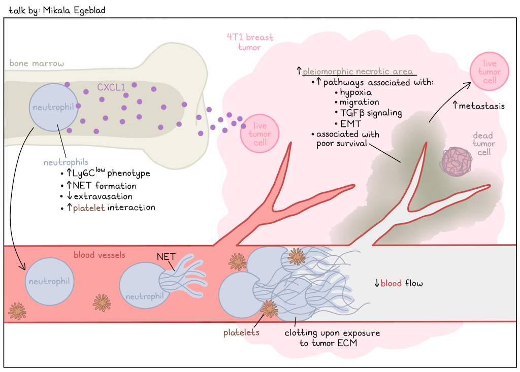
While neutrophils are the most abundant white blood cells in the human body and turn over rapidly (100 billion neutrophils are produced per day in humans), their study in cancer immunology has been limited. Neutrophils exhibit a unique type of cell death, forming neutrophil extracellular traps (NETs) that contain several components, including DNA, proteases, and cytokines. Mikala Egeblad discussed work on neutrophils and NETs in cancer. In 4T1 breast tumors, necrotic areas can be detected that are pleomorphic and contain NETs. Tumor necrosis has long been known to be associated with metastasis and early recurrence, and in a breast cancer dataset, these areas were associated with poor survival. In human tumors with necrosis, aggregates comprising neutrophils and NETs were detected close to the necrotic regions. This led Egeblad to hypothesize that these aggregates were blocking the bloodstream and causing necrosis in the downstream tissue supplied by these vessels. In the Lewis lung carcinoma model, necrotic areas did not form when neutrophils were absent. Several tumor models had this pleomorphic necrosis, but not all did. Comparing the neutrophil populations in mice with tumors that had this necrosis type and those that did not revealed a population of neutrophils with low expression of Ly6C in pleomorphic necrotic tumors. CXCL1 secreted by cancer cells was found to drive the generation of these Ly6Clow neutrophils and necrosis. These neutrophils were vasculature-restricted, with impaired extravasation, and were better at forming NETs. In mice, those with reduced NET formation through knockout of Pad4, encoding an enzyme critical for NET formation, had less tumor necrosis and fewer metastases. Perinecrotic regions exhibited increased expression of pathways associated with hypoxia, migration, TGFβ signaling, and epithelial-to-mesenchymal transition (EMT). In tumor cells with dominant-negative TGFBR2, there was a reduction in metastases, but not necrosis. Based on this, Egeblad described a model in which the tumor produces CXCL1 that alters neutrophil maturation in the bone marrow, resulting in a Ly6Clow population, which then traffics to the tumor and gets exposed to areas in the blood vessels where the extracellular matrix is exposed. Platelets also go to these areas, and a feed-forward loop can be observed between neutrophils and platelets, leading to clotting in the vasculature, and resulting in blockage and downstream necrosis. Further, these perinecrotic regions drive metastasis through increased TGFβ and EMT. Tumor necrosis may therefore be targeted by blocking NET formation. Egeblad also noted that about 20% of patients with late-stage cancer die from clotting abnormalities, and they are investigating whether these neutrophils are forming NETs in other places in the body, contributing to this mortality. This process might be relevant not just to cancer, but also to other diseases, such as COVID-19.
Reading suggestion:
Neutrophils not so neutral in tumors after all
Leukocyte Ig-like Receptor B (LILRB) checkpoints - Marco Colonna – Massachusetts General Hospital
The study of how macrophages balance the multitude of activating and inhibitory receptor signaling to determine function has been a long-standing interest of Marco Colonna. Based on his work, and that of others, there is now intense interest in suppressing the activating receptor TREM2 to remodel T cell suppressive macrophage to a more antitumor, T cell-activating state. However, in this talk, Colonna focused on the actions of the inhibitory leukocyte immunoglobulin-like receptor B family (LILRB). In humans, there is a family of five LILRB orthologs (1–5), which are co-localized on chromosome 19. In mice, however, there are only two orthologs, PIRB and Lilrb4a (aka GP49B1), which are located on different chromosomes; PIRB is in the corresponding chromosome location to the human LILRB complex. Human LILRB4 is found on multiple myeloid cells (monocytes, macrophages, cDCs, pDCs) and on some B cells, and there are four identified ligands: APOE, fibronectin, secretogranin 2, and galectin-8. LIILRB4-expressing macrophages can be readily detected in lung cancer, often colocalized with fibronectin-expressing cells, and there is a negative correlation between LILRB4 expression levels and survival in NSCLC. Two groups have shown preclinically that knockout of Lilrb4 or antibody blockade of Lilrb4a improved tumor control in different models (MC38 and metastatic LLC). However, linking the effects of LILRB in humans and mice is confounded by the differences in chromosomal location, and thus potential regulatory control differences. As examples of this complexity, Colonna pointed out that in the mouse, T cells, mast cells, and NK cells express Lilrb4a, and pointed to the work of Bob Schreiber, who showed that particular vaccine conditions in the MCA/1956 tumor model produce Lilrb4a+ regulatory TR1 T cells, suggesting a potentially complex, context-dependent regulation of Lilrb4a. To eliminate this as a variable, Colonna inserted the human LILRB gene locus as a BAC clone into the corresponding mouse chromosome (Telo Tg mice). In TeloTg mice, LILRB4 is expressed in similar tissues and cell types as in humans, but is no longer expressed in T cells, mast cells, or NK cells, recapitulating the human pattern. Administration of an anti-human LILRB4 antibody in three different s.c. tumor models (MC38, MCA/1956 and B16), however, failed to show an effect. To see if this was related to the absence of the proper ligand, the team performed immunohistochemistry for fibronectin, and observed that in s.c. MC38 tumors, fibronectin could be detected on the outside capsule of the tumor, while in B16F10 metastatic lesions in the lung, fibronectin was both outside and inside the tumor, and LILRB4+ macrophage could be observed in association with fibronectin inside the tumor. Blocking LILRB4 in the B16F10 lung metastasis model showed a reduction in the number of metastatic nodules, and LILRB4+ macrophages could be observed in association with fibronectin inside the tumor. The importance of ligand binding to altering macrophage properties also came out of studies of two different anti-human LILRB4 antibodies that are currently in clinical development. Despite similar specificity, binding potency, and ability to reach and infiltrate the tumor between the two antibodies, one antibody was effective in tumor control, while the other was not. In a reporter assay following fibronectin binding, the antibody active in tumor control blocked binding of LILRB4 to fibronectin, while the other did not. Both antibodies blocked the interaction between LILRB4 and its other ligands. Alpha-fold was used to determine the fibronectin binding site of LILRB4, and site-directed mutagenesis confirmed this assignment. Furthermore, the effective LILRB4 antibody interfered with this interface. The LILRB4 antibody increased the frequency and activation of macrophages, cDCs, and T cells in B16 lung metastases. scRNAseq of tumors treated with the functional antibody showed that macrophages, monocytes, and DCs acquired an interferon-stimulated gene signature in the tumor, marked by high CXCL9 expression, similar to the signature observed by Anna Obenauf in inflammatory monocytes capable of promoting CD4+ and CD8+ T cell activation and tumor control. Blockade of INFAR or elimination of macrophages with anti-CSF1R both eliminated the antitumor effect, indicating that anti-LILB4 antibody activity depends on the correct epitope specificity of the antibody operating in a tumor immune environment enriched in fibronectin and myeloid cells.
Reading suggestion:
New target can put the brakes on macrophage and Treg immune suppression
Cleavable albumin-hitchhiking STING agonists for innate immune activation and combination with adoptive cell therapy in solid tumors - Neil Chada, BS – Vanderbilt University
Presenting research from his dissertation, Neil Chada discussed leveraging the cGAS/STING pathway to enhance the cancer immunity cycle. To overcome the limitations of cyclic dinucleotides (which are hydrophilic and negatively changed) and non-cyclic dinucleotides (which distribute systemically and are rapidly cleared, requiring intratumoral administration) as STING agonists, Chada and colleagues developed an albumin-targeted nanobody (nAlb) and used the enzyme sortase to install an azide that enabled conjugation of various small-molecule payloads. When they conjugated their nALB with the non-cyclic STING agonist diABZI, the drug effectively accumulated in murine tumors (and to a lesser extent in some other organs). As a result, tumors showed increased antitumor gene signatures and increased infiltration by CD4+ and CD8+ T cells, many of which showed an activated (PD-1+, Ki67+) phenotype. Next, Chada evaluated the nALB–diABZI in a B16F10-OVA melanoma tumor model with adoptive transfer of OT-I cells. Here, 3 doses of nALB–diABZI after OT-1 transfer slowed tumor growth and extended survival in mice, with similar results in the YUMM-OVA model. Tumor evaluation after 1 dose showed that TAMs were reduced, and OVA peptide presentation and CD86 were increased on macrophages. OT-I cells also showed increased granzyme B expression, suggestive of enhanced antitumor immunity. To further enhance the adaptability of their nanobody–drug conjugates, Chada and colleagues developed a new conjugation strategy using two different peptide coils – one that was recombinantly installed on the nAlb, and another that would form heterodimers with the first. This strategy allows for nAlb to be conjugated with more possible payloads (particularly hydrophobic non-cyclic agonists), requires no specific recombinant enzyme, and results in higher product yields. While conjugates developed with this strategy showed shorter half-lives, they ultimately had similar distribution profiles, suggesting that this strategy could also be effective for making nanobody–drug conjugates for the preferential delivery of STING agonists or other small-molecule payloads to tumors.
Reading suggestion:
STING Licensing of Type I Dendritic Cells Potentiates Antitumor Immunity
Metabolism
Postprandial control of T cell immunity: implications for checkpoint blockade - Greg M. Delgoffe, PhD – University of Pittsburgh
Greg Delgoffe discussed the impact of meals on the immune response. While most T cells are in a more quiescent state, once activated, their metabolism increases to allow for proliferation, infiltration, and cytotoxicity. In memory responses, cells rewire mitochondria to increase respiratory capacity, while metabolism is limited in exhausted cells. Most research on the effects of diet on the immune response has been long-term. Instead, Delgoffe asked how a meal acutely impacts the immune system. Healthy volunteer blood was collected before breakfast after an overnight fast, and 6 hours later after breakfast (and lunch). With this, post-feeding differences in T cell metabolism could be detected. When these cells were expanded in vitro, the “fed” cells expanded more and produced higher levels of cytokines over the course of the culture than the “fasted” cells, so changes persisted after several rounds of cell division. No time-of-day effects were observed in human samples, and the microbiome was also not a confounder in mice. When fasted or fed T cells from OT-I TCR transgenic mice were transferred into fed mice infected with a vaccinia virus expressing OVA , the fed T cells performed better and, when isolated again after 11 days, still retained higher respiratory capacity and other metabolic features. Additionally, the fed T cells differentiated into a greater percentage of effector memory cells after 45 days in vivo. Delgoffe argued that the timing of T cell activation is important, with fed T cells outperforming fasted T cells when they encountered their corresponding antigen in the mouse right away. However, when the infection of the recipient mouse was delayed, this effect was gone. Hypothesizing that metabolites might be different, Delgoffe and team incubated T cells from the fasted condition with serum from the fed condition, and found that this recreated the “fed” phenotype. Meanwhile, fasted serum had no impact on fed T cells. Using various inhibitors, the researchers showed that fed T cells were sensitive to inhibitors of lipid metabolism, and lipids as the only nutrient source could induce the postprandial effect in mice. The transcriptome of T cells from fed mice did not differ from fasted mice, but their proteome did, particularly when cells were in an activated state. Routine instructions frequently direct donors and patients to fast prior to blood collection, and a fasted state might similarly skew immunological analyses performed on clinical or surgical tissue samples. To determine the consequences for CAR-T cells, fed and fasted human T cells were used for CAR-T manufacturing. The fed CAR-T cells had higher cytotoxicity in vitro. In vivo, the fasted cells were initially effective, but failed to persist, whereas the fed cells did persist, and demonstrated enhanced antitumor efficacy. With immune checkpoint blockade (ICB), Delgoffe noted that it is difficult to define the timing of T cell activation. Therefore, they assessed whether never entering a fasted state would improve efficacy. Using meal replacement drinks before sleep in mice improved the efficacy of ICB. In healthy volunteers, drinking meal replacement drinks before bed could prevent the fasted-state effects in T cells. These findings inspired a neoadjuvant clinical trial in melanoma (FASTBREAK) with either a daytime or a pre-nocturnal oral meal replacement given to patients during the neoadjuvant period.
Reading suggestion:
Stressed out telomeres drive T cell malfunction
By Ute Burkhardt, Ed Fritsch, Lauren Hitchings, and Maartje Wouters


