

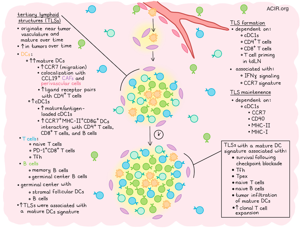
The presence of tertiary lymphoid structures (TLSs) in tumors is associated with improved outcomes and response to immune checkpoint blockade (ICB). Although the cell populations within these TLSs are well established, the precise contributions of DCs to TLS formation and maintenance remain largely unknown. Mattiuz et al. investigated this question using human tissue samples and a non-small cell lung cancer (NSCLC) murine model that spontaneously forms TLSs. Their results were recently published in Science.
The researchers analyzed human NSCLC, HCC, colorectal cancer, and renal cell carcinoma using spatial transcriptomics. They found intratumoral TLS areas enriched with mature DC, cDC1, Tfh cell, PD-1+CD8+ T cell, naive T cell, memory B cell, and germinal center B cell molecular programs. Mature DCs were significantly more abundant in TLSs than cDC1s and cDC2s, with cDC1s expressing more mature DC genes than cDC2s, indicating that tumor antigen-loaded cDC1s accumulate in TLSs, as DC maturation is linked to cellular debris uptake. Multiplex imaging confirmed these findings, showing DC-LAMP+ mature DCs and CLEC9A+ cDC1s enriched in TLSs.
In ICB-treated patients, pretreatment biopsies of NSCLC and metastatic kidney cancer with higher TLS density expressed the mature DC gene signature and were associated with improved survival. The mature DC signature correlated with signatures of Tfh cells, Tpex cells, and naive B and T cells. Further, tumor resections of HCC obtained after treatment with neoadjuvant ICB showed that patients with higher numbers of TLSs had improved disease-free survival, increased tumor infiltration of mature DCs, and T cell clonal expansion.
To assess the mechanistic role of DCs in TLS formation and function, the researchers moved to a murine model in which the lung adenocarcinoma cell line KP-HELLO-2 was inoculated intravenously, resulting in orthotopic tumor lesions with high numbers of TLSs. These TLSs had distinct T and B cell zones, containing germinal centers with stromal follicular DCs and B cells. CCR7+MHC-II+CD86+ DCs accumulated in the TLSs and interacted with CD4+ and CD8+ T cells and B cells based on immunohistochemistry.
To assess if cDCs influence TLS formation, the model system was used to assess whether removing cross-presenting cDC1s affected TLSs. Without cDC1s, tumors had fewer and smaller TLSs, fewer Tfh, IFNγ+CD4+ T, CD8+ T cells, and germinal center B cells, and higher tumor burden. Depleting CD4+ or CD8+ T cells also reduced TLSs. Upregulation of IFNγ signaling and CCR7 gene signatures in cDCs were associated with TLS formation; tumor infiltration of Tfh cells, IFNγ+CD4+ T cells, CD8+ T cells, and germinal B cells; and lower tumor burden.
The researchers used FTY720 to inhibit lymphocyte egress from TDLNs to determine if TLS formation depends on cDCs at the tumor site or in TDLNs. Inhibiting T cell egress in the first 9 days post-inoculation reduced TLS formation; treatment after day 9 had limited effect. Therefore, T cell priming in the TDLN seems required for TLS formation, but not maintenance. Longitudinal profiling showed immature TLSs originate near tumor vasculature (based on spatial transcriptomics and multiplex IHC) and mature over time. TLS number and size increased during tumor progression, especially after day 8. The number of mature cDC1s and cDC2s with tumor antigens rose until day 8 in the TDLN, then declined and accumulated in tumors. CD4+ populations remained stable, while antigen-experienced and exhausted CD8+ T cells increased during tumor progression. At later time points, there was expansion of Tpex cells, germinal center B cells, and tumor-specific IgG antibodies.
To establish the role of cDCs in TLS maintenance, two models were generated in which either the entire cDC compartment or only cDC1s were depleted starting at day 8 after tumor inoculation. Both strategies reduced TLS numbers. cDC1 depletion resulted in reduced proportions of tumoral Tfh cells, leading to disappearance of germinal centers, a decrease in CD8+ T cells and proliferating cells in the TLS, as well as a reduction in germinal center B cells and tumor-specific IgM and class-switched IgG antibodies. Since this suggests that cDC1s are required locally for TLS maintenance and function, the researchers then assessed blocking late-stage TDLN egress at day 8. While this did not impact the number of TLSs, concomitant cDC1 depletion at day 9 reduced TLS maintenance, while administration of FLT3L increased the number of cDCs and TLSs.
To examine the molecular signals that drive cDC migration and support TLS maintenance, the researchers created a ligand–receptor analysis map. Mature DCs in TLSs expressed the highest levels of ligand-receptor pairs interacting with CD4+ T cells. Mature DCs expressed high levels of CCR7, and perivascular cells and cancer-associated fibroblasts (CAFs), expressing its ligand CCL19, were enriched in TLSs near mature DCs. To determine whether deletion of CCR7 in cDC1s after T cell priming and tumor recruitment compromises TLS maintenance, the researchers created a model with temporal deletion of CCR7 in the cDC1 subset at day 8 after tumor engraftment. This reduced the number and size of TLSs and reduced the number of Tfh cells in tumors. CCR7 deletion from the whole cDC compartment gave similar results, while conditional CCL19 deletion from cDC1s did not impact TLS maintenance. Therefore, cDC1 migration to CCR7 ligand-enriched stromal hubs seems required for TLS maintenance.
cDC1s are known to recruit T cells via a process requiring CD40 signaling. Depletion of CD40+ cDC1s at day 8 after tumor implantation reduced the number and size of TLSs and CD8+ T cell infiltration. When MHC-II was deleted from cDC1s at day 8, the number and size of TLSs also reduced, with accompanying decreases in Tfh, CD8+, and PD1+CD8+ T cells, as well as tumor-binding IgG antibodies, and increases in Tpex cells. Interestingly, deletion of MHC II could not be rescued by CD40 agonism, indicating a direct role for antigen presentation by MHC II+ DCs. MHC-I depletion in a similar setting also reduced the number and size of TLSs, but the effect was weaker than MHC-II depletion.
Together, these data suggest that cDC1s play essential roles in the formation and maintenance of TLSs in tumors. These data may provide clues on how to strategically induce and maintain TLSs in patient tumors to improve immunotherapy outcomes.
Write-up by Maartje Wouters, image by Lauren Hitchings
Meet the researcher
This week, lead author Raphaël Mattiuz answered our questions.

What was the most surprising finding of this study for you?
One of the biggest open questions in the field has been understanding which immune cells organize protective immune hubs (tertiary lymphoid structures) inside tumors – how they form initially and how they are maintained over time. What surprised us most was that this role is driven by one of the rarest immune cell types in the body: type 1 dendritic cells. Despite their scarcity, they act as the conductors of the antitumor response. They first help build these immune hubs, and then relocate into them, continuously coordinating T cells and B cells to sustain local anti-cancer activity. Remarkably, they orchestrate both cancer-killing T cells and antitumor antibody responses, bridging the two main arms of antitumor immunity directly inside the tumor.
What is the outlook?
Our findings identify dendritic cells as a promising target for the next generation of cancer immunotherapies. If we can safely increase their numbers or enhance their activity, we may be able to promote the formation of these protective immune hubs and make antitumor immune responses stronger and more durable. Our next goal is also to understand how these immune hubs generate long-lasting immune memory that prevents cancer from returning after treatment.
If you could go back in time and give your early-career self one piece of advice for navigating a scientific career, what would it be?
I would tell myself to trust my curiosity and follow the questions that genuinely excite me. I became fascinated by these immune hubs nearly ten years ago after reading papers describing similar structures during infections. I kept wondering whether dendritic cells in these hubs played a similar role in cancer, and that question stayed with me throughout my training. It took almost a decade before I finally had the opportunity to answer it. Looking back, my advice is simple: if a scientific question keeps coming back to you, follow your gut and pursue it.


