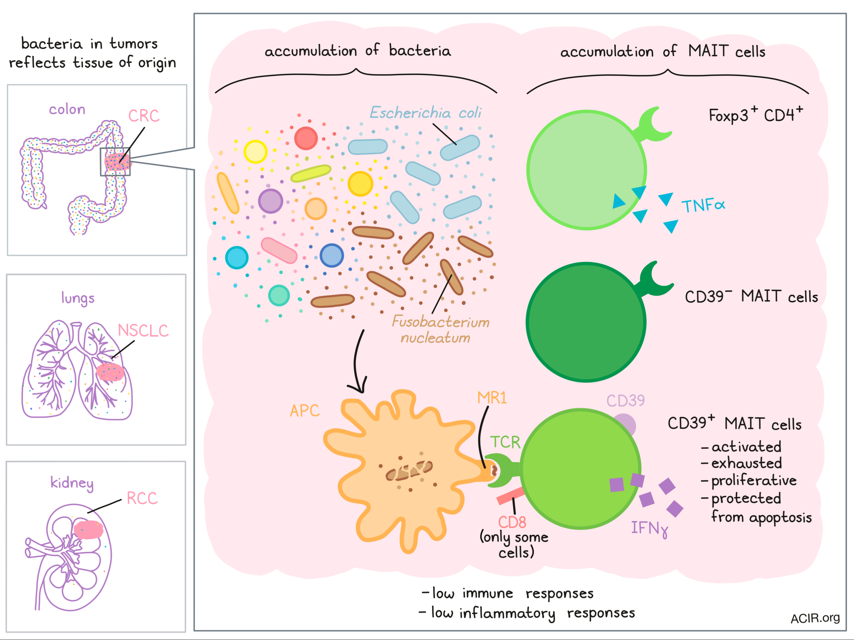
In patients with colon adenocarcinoma, high infiltration by mucosa-associated invariant T (MAIT) cells – which can be activated by a wide variety of bacteria and recognize epitopes presented by the MHC-like molecule using a limited set of invariant/preferential TCRs – have recently been associated with poor clinical outcomes. In a study recently published in Cell Reports Medicine (a new journal from Cell Press), Li et al. investigated the role of MAIT cells in cancer, shedding new light on how the microbiome impacts antitumor immune responses.
Based on evidence from prior studies, Li et al. investigated the hypothesis that MAIT cells can be activated by and respond to gut microbiome-associated antigens within the tumor microenvironment. To begin, they profiled samples from colorectal cancer (CRC), non-small cell lung carcinoma (NSCLC), and renal cell carcinoma (RCC) and found that MAIT cells accounted for a higher portion of total T cells in CRC compared to NSCLC or RCC. As no such difference was observed in the periphery, this suggested a tumor-specific accumulation of MAIT cells in CRC.
Comparing the profiles of MAIT cells within CRC to MAIT cells from adjacent tissue or PBMCs, the researchers identified a tumor-specific phenotype characterized by a distinct transcriptomic profile (enrichment for TCR signaling and negative apoptotic regulation pathways), high expression of CD69, CD103, CD38, and CD39, and lower expression of CD27 and CD49d compared with peripheral MAIT cells. Most tumor-infiltrating MAIT cells also expressed markers indicative of response to inflammation. Interestingly, the researchers also observed heterogeneity within tumor-infiltrating MAIT cells from CRC that was not observed in the periphery.
Among tumor-infiltrating MAIT cells, Li et al. identified a distinct cluster of CD4+ cells, some of which surprisingly co-expressed CTLA-4, Foxp3, and other markers that are typically associated with Tregs. Functional analyses revealed that unlike Tregs, CD4+Foxp3+ MAIT cells produced the pro-inflammatory cytokine TNFα, suggesting that Foxp3 expression on CD4+ MAIT cells might be a marker of activation rather than a marker of immunosuppressive function. The researchers also identified distinct signatures between CD39+ and CD39- MAIT cell populations. Further, within CD39+ MAIT cells, there was one subpopulation that expressed CD69 and CD103, both markers of tissue residency, along with CD38, and another that expressed CTLA-4, Tim3, GITR, and CD45RA.
Looking more closely at MAIT cells expressing CD39, Li et al. found that the proportion of CD39+ MAIT cells was increased in CRC, with many of these cells co-expressing CD8. The CD39+CD8+ MAIT cell population also frequently expressed markers of tissue residency and genes associated with activation and exhaustion. Further, many CD39+ MAIT cells showed evidence of increased proliferation and enhanced protection against cell death compared to CD39- MAIT cells. While CD39+ MAIT cells showed little evidence of polyfunctionality, most were able to express Granzyme A, Granzyme B, or perforin. Together, this evidence suggested that tumor-infiltrating CD39+ MAIT cells might represent a population of MAIT cells that had been activated in response to antigen stimulation.
To test the functional responses of MAIT cells, Li et al. showed that stimulation of MAIT cells with Escherichia coli induced expression of CD39. Blocking stimulation with an anti-MR1 antibody abrogated this effect, indicating that CD39 expression was induced by TCR-driven activation. Based on these results, the researchers hypothesized that the phenotypes and functions of tumor-infiltrating MAIT cells could be affected by bacteria and/or metabolites present in the tumor microenvironment in CRC. Consistent with CD39 expression as a marker of antigen stimulation, they observed that CD39 expression was higher in CRC compared to NSCLC or RCC, reflective of the abundance of bacteria typically found in the colon relative to lungs and kidneys. Furthermore, analysis of CRC tumors showed that that the bacterial load was higher in tumors than in paired adjacent tissue, as was the expression of infiltrating CD39+ MAIT cells. Further, expression of CD39 on MAIT cells was particularly enhanced in tumors with high bacterial loads compared to lower bacterial loads, suggesting that when bacteria was more abundant, MAIT cells were more likely to become activated and express CD39. Other than CD39, the expression profile of MAIT cells was fairly consistent across CRC, NSCLC, and RCC, further indicating that the unique CD39 expression on tumor-infiltrating MAIT cells in CRC occurs as the result of antigen exposure.
Between patients, bacterial loads were heterogeneous, though bacteria from the same phylum were generally correlated. Most bacterial strains were not particularly abundant, though a few, including E. coli, were notably enriched. To determine whether certain gut bacterial strains associated with CRC could affect the functions of MAIT cells, Li et al. selected a few strains of bacteroides and fusobacterium that were highly abundant in CRC. In in vitro stimulation assays, only Fusobacterium nucleatum or culture supernatents from F. nucleatum activated MAIT cells and stimulated production of IFNγ – an effect that was dependent on TCR engagement.
Exploring whether MAIT cells might impact patient clinical outcomes, Li et al. found that patients with CRC that was highly infiltrated by bacteria showed evidence of low immune and inflammatory responses. Within these patients, MAIT cells expressed markers of wound healing and tissue repair that would suggest a role in promoting epithelial growth. The gene for PyrD, an enzyme that is involved in riboflavin synthesis and supports MAIT cell development and activation, was enriched in metagenomes from CRC versus normal colon samples, suggesting that tumor-specific characteristics might play a role in bacterial infiltration and thus MAIT cell activation. Evaluating the frequency of MAIT cells across clinical stages revealed a trend, not reaching statistical significance, of higher frequency and later stage.
Overall, the results uncovered by Li et al. provide evidence that MAIT cells in CRC can be activated by and respond to microbial antigens in a TCR-dependent manner. This activation appears to be marked by the expression of CD39. Given results that the presence of MAIT cells seems to affect clinical outcomes in patients with CRC, this early research could pave the way for cancer immunotherapies that target MAIT cells or the microbiome.
by Lauren Hitchings
Meet the researcher
This week, Shamin Li and Evan Newell answered our questions.
What prompted you to tackle this research question?
SL: As my PhD focused on NKT cells, I was familiar with unconventional T cells and was interested in MAIT cells, which were still a not-so-well documented immune population, especially in the context of human malignancies. Since the research on interrelationships between cancer therapies and the microbiome was booming, I took the opportunity of our large cohort of human colorectal cancer samples to assess MAIT cell infiltration in the tumors. We were hoping that these cells recognizing bacterial metabolites could be somehow connected to bacterial infiltration in tumors, and we were able to find evidence that indicated the importance of MAIT cells in microbiota-associated cancers.
EN: I’m generally interested in how T cell antigen specificity can give rise to a multitude of different T cell populations with different functions, and then how they can work together to provide immunity to cancer and infectious disease. Although their roles are still mysterious, MAIT cells are a nice example of a highly abundant (especially in human blood and other tissues) population of T cells for which the antigen specificity is known, but roles in disease are much less understood. This was definitely encouraging when it came to Shamin’s ambition to ask more about how these cells might contribute to cancer immunity.
What was the most surprising finding of this study for you?
SL: As CD39 is a marker associated with chronic TCR activation on conventional T cells, we were intrigued by the high expression of CD39 on tumor-infiltrating MAIT cells that was only found in colorectal cancer. We hypothesized that induction of this expression could be initiated by bacterial antigen recognition, an idea that we confirmed next by in vitro stimulation assays. The observation of a dominant CD39+ MAIT cell population in colorectal cancer is a key to this work, and the presence of a Foxp3+ subset found within it could be another interesting puzzle to solve.
EN: Because MAIT cells are known to be able to functionally respond to non-specific stimulation such as cytokines, I expected that MAIT cells would not necessarily show signs of active TCR-driven responses in cancer. So, it was surprising to find that tumor-infiltrating MAIT cells expressed markers associated with T cell exhaustion, including CD39, which we are hypothesizing to predominantly require a TCR-driven signal for its expression. I was also surprised to see Foxp3-expressing CD4+ MAIT cells in human tumors.
What was the coolest thing you’ve learned (about) recently outside of work?
SL: During my break times, I enjoy watching wildlife documentaries and reading about zoology, because I find animal life fascinating. The other day I was reading about how the auditory cortex of macaques works differently as compared to humans, and so playing a harmonic or a pitchless/noisy tune would sound the same to them. For someone who loves music, I was stunned that such a similar species to us could have such a different auditory neural response. It makes me realize how sometimes we take our perceptions for granted and reminds me that we should always take a step back to gain a different perspective.
EN: With young kids at home and in the months since the development of the COVID-19 pandemic, it has been both a source of stress and a real joy to spend more time at home than ever before. In trying to maintain their learning, I’ve particularly enjoyed reading a variety of books that I hadn’t had a chance to read before, as well as many from my own childhood. In thinking of how we might be able to improve our learning, I’m intrigued by an article I read recently suggesting that you can learn more efficiently by increasingly spacing the time between repetition. I’ll have to figure out how to implement this on my kids and the words we’ve learned – maybe there’s an iPad app for it?


