NK cells are promising targets for cancer immunotherapy, but clinical results have so far been limited. Therefore, there is a need to better understand NK cell subphenotypes and their function in tumors. Two recent papers characterized tumoral NK cells. In Cancer Immunology Research, Lozada et al. characterized proinflammatory and suppressive NK cells across tumor types. In Science Translational Medicine, Horowitz, Mohammad, Ho Shin, et al., characterized tissue-resident NK cells (trNK) and uncovered a highly cytotoxic trNK subpopulation.
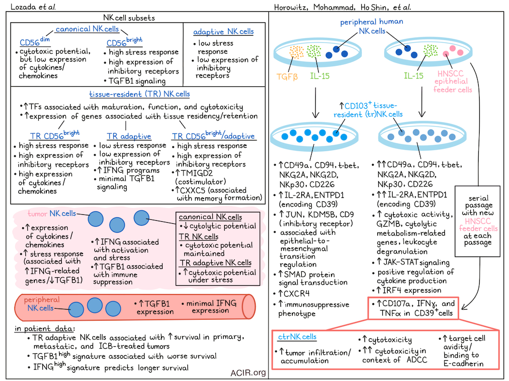
Lozada et al. started with clustering of NK cell subsets from a published scRNAseq dataset from 339 samples across 14 solid tumor types. There was heterogeneity within human NK cells, and two unique NK cell subsets of interest were identified: a tissue-resident (TR) adaptive NK cell subset and a TR subset co-expressing features of CD56bright and adaptive NK cells. Furthermore, well-known subsets, including canonical CD56dim and CD56bright subsets, and adaptive and TR phenotypes, were detected.
Assessment of gene signatures showed that canonical CD56dim cells had cytolytic potential, but expressed low levels of chemokines and cytokines. TR subsets had high levels of chemokine and cytokine expression. Adaptive and TR adaptive NK cells had the lowest stress response scores and inhibitory receptor expression levels, while canonical CD56bright and TR CD56bright expressed the highest levels of both.
Compared with their conventional counterparts, TR cells highly expressed genes associated with tissue residency and retention, and upregulated transcription factors involved in NK cell maturation, function, and cytotoxicity. TR CD56bright/adaptive NK cells upregulated TMIGD2, encoding a costimulatory immune receptor, and CXXC5, encoding a transcription factor associated with memory formation in NK cells.
Comparing the NK cell compartment across cancer types showed clear differences in proportions of TR and conventional subsets between tumors. Comparing NK cells in the periphery with those in the tumor showed consistent upregulation of chemokines and cytokines and stress response genes in the tumor. While conventional subsets had reduced cytotoxic potential in tumors, most TR subsets maintained their immunomodulatory activity.
In several cancer types, tumor-infiltrating NK cells had higher stress-response signatures. However, NK cells in high-stress tumors maintained expression of cytotoxic and immunomodulatory genes. Stress was associated with upregulation of IFNG-related genes, which may reflect an activated cell state, and TGFB1 expression was negatively correlated with IFNG. While several subsets showed reduced cytotoxicity under higher stress, adaptive NK cells showed increased cytotoxicity under stress.
The researchers then assessed whether TGFB1 and IFNG define the different functional states. Most NK cells in the periphery expressed TGFB1 and lacked IFNG, while most NK cells in the tumor expressed only IFNG. IFNGhigh NK cells upregulated activation and stress markers, while a TGFB1 signature was correlated with immunosuppression. Canonical CD56bright NK cells had more TGFB1 signaling, while TR adaptive NK cells had mostly IFNG programs and minimal TGFB1 involvement.
To assess the clinical impact of the NK cell phenotypes, transcriptional signatures for all six populations were projected onto a pan-cancer cohort of bulk RNAseq samples from 53 cancer types. High levels of TR adaptive NK cells in tumors were associated with improved overall survival across cancers, while CD56bright NK cell subsets had a modest beneficial effect. TR adaptive NK cells were also the most favorable subpopulation in the context of ICB treatment.
In primary tumors, only canonical CD56dim and TR adaptive NK cells predicted better outcomes, while in metastatic tumors, all adaptive or TR subsets had favorable effects. TR adaptive NK cells were the only subset associated with better outcomes in primary, metastatic, and ICB settings. The frequency of TGFB1-signaturehigh NK cells correlated with worse survival, while IFNG-signaturehigh NK cells predicted longer survival.
In the second study, Horowitz, Mohammad, Ho Shin, et al. showed that two in vitro methods could differentiate peripheral human NK cells into tissue-resident (tr)NK cells. Exposing NK cells to either head and neck squamous cell carcinoma (HNSCC) epithelial feeder cells plus IL-15 or to TGFβ plus IL-15 induced CD49a and CD103 expression. Comparing these induced trNK cells showed that CD103+ NK cells from HNSCC/IL-15 culture conditions separated from the ones obtained with TGFβ/IL-15 cultures. The HNSCC-induced cells expressed higher levels of CD49a, CD94, t-bet, NKG2A, NKG2D, NKp30, and CD226, and showed greater cytolytic activity against target cells.
scRNAseq of these trNK cells showed that the HNSCC-induced trNK cells predominantly expressed the GZMA cytolytic gene, some metabolism-related genes, and higher levels of IL2RA and ENTPD1 (encoding CD39) than the TGFβ-induced variants. The TGFβ-induced trNK cells had higher expression of JUN and KDM5B, as well as CD9, an inhibitory receptor. The HNSCC-induced cells were enriched for pathways related to leukocyte degranulation, receptor signaling via the JAK–STAT pathway, and had positive regulation of cytokine production, while the TGFβ-induced cells were associated with epithelial-to-mesenchymal transition regulation and SMAD protein signal transduction.
A correlation analysis between DEGs and differentially accessible regions identified by ATACseq revealed that IRF4, previously associated with increased NK cell expansion, was highly expressed in HNSCC-induced trNK cells. TGFβ-induced cells showed increased CD9 and CXCR4 expression and chromatin accessibility, resembling the immune-regulatory decidual NK cell subtype.
CD49a+CD103+ NK cells in human primary HNSCC tumors showed heterogeneity in their expression of CD39. Further, TCGA data of HNSCC showed a positive correlation between ENTPD1 (CD39) and NK cell cytotoxic molecules, while it negatively correlated with CD9 expression. In vitro stimulation of HNSCC-induced trNK cells showed that CD39+ cells had higher levels of the degranulation marker CD107a and more IFNγ and TNFα. The researchers named this population cytotoxic trNK (ctrNK) cells.
ctrNK cells had significantly higher cytolytic activity in vitro, and their cytolytic activity increased even further in the context of antibody-dependent cellular cytotoxicity (ADCC). CAR-NK cells grown in the presence of IL-15 and HNSCC cells showed enhanced killing capacity.
To assess tumor infiltration capacity, the cells were cocultured with spheroids derived from HNSCC cell lines. ctrNK cells showed the best infiltration and accumulation in these spheroids, dependent on CD103 signaling. ctrNK cells had higher avidity for target cells and binding to E-cadherin than control NK cells. Treatment of mice bearing solid HNSCC tumors with adoptive transfer of ctrNK cells resulted in higher CD49a+CD103+ NK cell tumor infiltration.
Expanding on the adoptive transfer data, the researchers established a ctrNK cell enrichment and expansion method using serial passage of ex vivo-differentiated ctrNK cells, with new HNSCC feeder cells at each passage. Adoptive transfer of the obtained ctrNK cells to three mouse tumor models resulted in reduced tumor burden. When this treatment was combined with cetuximab in an EGFR+ HNSCC tumor model, the combination better controlled tumor formation than either monotreatment.
Overall, the data in these two studies show the importance of tissue-resident NK cells in tumors, and the specific transcriptomic and phenotypic features that impact antitumor responses. Modulation of these subtypes via immunotherapy may enhance the efficacy of NK cell-based immunotherapy approaches. Further work to consolidate the description of these subsets of tumor-resident NK cells will further progress in the utilization of NK cells for immunotherapy.
Write-up by Maartje Wouters, image by Lauren Hitchings
Meet the researcher
This week, John Roy Lozada, first author on “Integrated Single-Cell Profiling Reveals Dichotomous NK Cell Populations Associated with Immunosuppression in Solid Tumors” and Nina Horowitz, first author on “CD39+CD49a+CD103+ cytotoxic tissue-resident natural killer cells infiltrate and control solid epithelial tumor growth in mice” answered our questions.
What was the most surprising finding of this study for you?
JRL: We uncovered an underappreciated, dichotomous role for NK cells in promoting versus restraining antitumor immune responses through contradicting cytokine programs. Intriguingly, this was also linked to their phenotypic profiles, which vary based on tumor context.
NH: The impact of the phenotypic shift on tumoroid infiltration rates really surprised us. We were expecting to see some improvement with the novel phenotype, of course, but the magnitude of the change was quite dramatic. The tumoroids that were co-cultured with the tissue-resident NK cells absolutely lit up under the microscope! Dr. Sunwoo and I were both so skeptical that we immediately repeated the experiment to be sure the results were real! Fortunately, the following experiments showed that the effect was significant, consistent across donors, and reproducible.
What is the outlook?
JRL: We are hopeful that our work will provide a foundational framework to understand both the phenotypic and functional diversity of NK cells across cancer, and illuminate strategies to enhance the NK cell-mediated response against cancer. This can be achieved through multiple avenues suggested by our study, including harnessing specific adaptive and tissue-resident populations of NK cells imbued with superior antitumor properties, and by skewing the balance of functional programs towards IFNγ-driven pro-inflammatory cascades.
NH: Our next challenge is to manufacture these cells in a scalable manner so they can be tested for patients with solid tumors! If successful, I think this could have a massive impact on the field of solid tumor immunotherapy. In the same way that TIL therapies are a significant improvement to conventional CAR T cell therapies, these novel tissue-resident NK cell therapies could exhibit increased efficacy when compared to prior CAR NK attempts. I believe we have ignored the NK phenotype question for too long, and I hope this data influences the perspective of the field.
If you could go back in time and give your early-career self one piece of advice for navigating a scientific career, what would it be?
JRL: Savor those early moments where you have freedom to explore and make mistakes. This goes hand in hand with finding the right mentors – those who support you, challenge you, and give you the space to find your own interests and pave your own path in academia.
NH: Don’t give up! It sounds so clichéd, but my career path had countless setbacks and struggles. If I hadn’t continued pushing forward time and time again, I wouldn’t be where I am today. A little-known fact is that the first time I applied to PhD programs, I was rejected from all of them outright – I didn’t even get invited for an interview! Luckily, I joined a program that let me take additional classes, and then two years later I re-applied. Thanks to that persistence, I was accepted to Stanford, graduated six years later, and now I have the good fortune of running my own cell therapy company called ImmuneBridge.


