

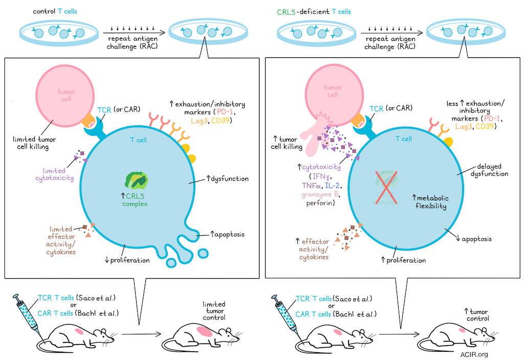
The lack of persistence after adoptive T cell therapies due to T cells becoming dysfunctional under chronic exposure to tumor antigens limits their efficacy. Two recent back-to-back publications by Bachl et al. and Saco et al. in Cancer Immunology Research aimed to address this by investigating functional knockouts in T cells under chronic tumor stimulation. Both studies found that disruption of Cullin RING ligase complexes led to T cell persistence and sustained tumor killing.
Bachl et al. conducted a genome-wide CRISPR-Cas9 knockout screen in primary human T cells, repeatedly stimulating them with antigen-matched cancer cells to model the exhaustion-inducing tumor microenvironment. T cells were transduced with an NY-ESO-1-specific TCR and a genome-wide sgRNA library, followed by Cas9 editing. Cells then underwent repetitive antigen challenge (RAC) with NY-ESO-1+ tumor cells every 48-72 hours. After nine rounds (RAC9), the TCR-T cells could no longer clear tumor cells. Next-generation sequencing of baseline and final co-culture samples identified knockouts enriched during the screen.
A STRING-based protein–protein interaction network analysis of the top 50 hits identified seven significantly enriched members of the Cullin-RING Ligase 5 (CRL5) complex. This complex consists of the scaffold protein cullin-5 (CUL5), the RING protein RBX2 (RNF7), the adaptor proteins Elongin B/C, and one of many substrate receptor proteins recognized by the CRL5 complex.
Similarly, Saco et al. conducted a pooled genome-wide CRISPR KO screen in primary human CD8+ T cells engineered with an NY-ESO-1-specific TCR and subjected the cells to RAC with HLA-matched, NY-ESO-I+ M407 melanoma cells. After each round, they isolated 4-1BB-expressing cells and quantified sgRNA abundance. Later RAC rounds (4–6) enriched several Cullin-RING E3 ligase components, with the CRL2 substrate receptor and tumor suppressor VHL as the strongest hit. CRL5 components CUL5, RNF7, and ELOB were also strongly enriched. A validation screen of 225 selected genes confirmed these as the most positively selected after later RACs.
Saco et al. validated the functional consequences of disrupting the detected components of the CRL2-VHL and CRL5 complexes over several RACs. Knockouts of CRL5 components led to enhanced T cell cytotoxicity (sustained through RAC8), with increased levels of IFNγ, TNFα, IL-2, granzyme B, and perforin. Bulk RNAseq after RAC1 and RAC3 showed that CRL5-deficient T cells clustered together and were distinct from control and VHL-deficient cells. After RAC1, CRL5-deficient T cells exhibited a transcriptional profile associated with increased effector activity and delayed dysfunction, with elevated levels of cytotoxicity-associated genes, effector cytokines, and effector-promoting transcription factors, and reduced expression of pro-apoptotic genes. These cells also showed reduced expression of inhibitory and exhaustion-related genes. These effects became more pronounced after RAC3.
Similarly, to assess the CRL5 complex’s role in T cell fitness, Bachl et al. individually knocked out highly ranked complex members and subjected the cells to four RACs. After RAC1, KO TCR-T cells showed slightly greater tumor killing than controls; this effect became pronounced after RAC4. At this later stage, all subunit KOs then produced more IFNγ and TNF, with CUL5 KO cells showing the highest levels. Lowering IL-2 supplementation during culture (to induce stress) had little early effect, but after RAC4, ARIH2 and CUL5 KO cells showed greater antitumor activity than controls, which was further enhanced by RAC6, at which stage only CUL5 KO cells achieved complete tumor clearance.
After RAC6, CUL5 KO TCR-T cells showed higher levels of type 1 (IL-2, IFNγ, TNF) and type 2 (IL-4, IL-5, IL-13) effector cytokines, proliferated more, and had lower percentages of PD-1+, CD39+, and LAG3+ cells than controls, suggesting preserved function under chronic stimulation. KO cells remained dependent on exogenous cytokines and responded to tumors only in an antigen-specific manner, which are important safety requirements.
Since one substrate receptor of CUL5 is CISH, which is known to enhance T cell antitumor activity, Bachl et al. compared CUL5 KO and CISH KO cells to determine whether the CUL5 KO effects were simply attributable to CISH. However, after RAC6, the CUL5 KO T cells outperformed the CISH KO T cells. Comparing individual KOs of CRL5 substrate receptors showed that CUL5 KO T cells exhibited the strongest antitumor effects, suggesting that the CUL5 KO reflects a broader loss of multiple substrate receptor activities.
To identify CUL5-dependent substrates beyond the JAK/STAT axis, Bachl et al. performed quantitative diGly (K-ε-GG) ubiquitylation proteomics on CUL5 KO TCR-T cells under resting, acutely stimulated (one restimulation with CD3/CD28), and chronically stimulated (RAC3) conditions. In the CUL5 KO cells, the strongest signals came from nutrient transporters and a second cluster comprising membrane trafficking and structural regulators.
To determine shared transcriptional programs among the various KOs, Saco et al. performed a combined analysis of ARIH2-, CUL5-, and RNF7-deficient T cells and compared these to controls. After RAC1, cell cycle progression and proliferation pathways were enriched, along with activation of oxidative phosphorylation and glycolytic pathways, consistent with a metabolically flexible state.
Both publications then assessed how these findings could be used therapeutically. Bachl et al. started with knockout of CUL5 in CD19-CAR-T cells. Similar to previous experiments, RAC with CD19+ A375 cells resulted in enhanced tumor killing of CUL5 KO CAR-T cells in vitro. Using another target antigen (BCMA or HER2), and/or either a CD28 or 4-1BB costimulatory domain or different scFv for the CAR-T constructs did not impact the effects of CUL5 ablation.
To investigate the in vivo impact, a preclinical CD19 CAR-T cell model was used in which NSG mice were engrafted with NALM-6 leukemia cells and treated four days later with two low doses of either CUL5 KO or control CD19-targeting CAR-T cells. CUL5 KO CAR-T showed a trend toward improved tumor control over control CAR T cells, and no main differences were detected in animal weight, suggesting therapy was tolerated.
Next, NSG mice engrafted with OPM2 multiple myeloma cells were treated with low dose BCMA-targeting CUL5 KO or control CAR-T. In this aggressive model, the CUL5 KO CAR-T showed a small improvement in tumor control. Given that the observed in vitro effects emerged after repeated stimulation, the researchers then used the same model and subjected the mice to five rounds of tumor rechallenge starting at day 55. At day 87, CAR-T cells were isolated from the bone marrow and spleen for analysis. In the bone marrow, the CUL5 KO CAR-T cells were enriched compared to controls, with no major difference observed in the spleen. Ex vivo killing assays showed higher killing capacity of the CUL5 KO T cells than controls. No significant differences were observed in terms of frequencies of naive, central memory, effector memory, or terminally differentiated effector memory T cell subsets in the bone marrow of mice treated with each CAR-T therapy.
Finally, the researchers asked whether the limited effects observed in vivo in the initial studies might also be due to the lack of human cytokines in NSG mice, limiting initiation of JAK/STAT signaling. To determine whether directly controlling local cytokine delivery could synergize with CUL5 KO, RAC assays were performed with supplementation of IL-2, IL-12, IL-15, IL-21, or IL7/IL-15. IL-2 supplementation resulted in the most optimal tumor control. Based on this, primary human T cells were engineered with an NY-ESO-1 TCR and a CD19-specific synthetic intramembrane proteolysis receptor (SNIPR) to drive IL-2 expression locally. Cells were subjected to RAC with CD19+ A375 cells, which showed that IL-2 worked synergistically with CUL5 KO.
Similarly, Saco et al. transferred gene-modified NY-ESO-1-specific TCR-T cells into immunodeficient mice bearing HLA- and antigen-matched A375 human melanoma xenografts. T cells lacking CRL5 components showed improved tumor control, particularly those deficient in CUL5 or RNF7. Mice receiving control T cells exhibited rapid tumor progression and reached endpoints by day 22, whereas a subset of mice treated with CRL5 component-deficient T cells achieved complete or partial remission.
The researchers also tested pharmacological inhibition of neddylation, which is required for all Cullin-RING E3 ligase activity. MLN4924, a selective inhibitor of the NEDD8-activating enzyme, improved tumor control and cytotoxicity in control T cells across multiple RACs. It had little effect on the already high cytotoxicity of CRL5-deficient T cells.
The data from these two studies suggest that disruption of the CRL5 complex in T cells promotes their persistence and sustained tumor killing capacity in chronic stimulation environments like tumors. These findings can be used to engineer improved T cell-based therapies for more durable control of solid tumors.
Write-up by Maartje Wouters, image by Lauren Hitchings
Meet the researcher
This week, first author on “Disruption of the CRL5 ubiquitin ligase complex enhances 1 engineered T cell 2 persistence under chronic tumor antigen exposure” Stephanie Bachl answered our questions.
What was the most surprising finding of this study for you?
One of the bigger challenges has been figuring out how to best translate the biology we uncovered in vitro into an in vivo setting. T cell behavior is strongly influenced by interactions with the surrounding environment, so moving human primary T cells from a controlled in vitro system into mouse models introduces an additional layer of biological complexity. This has also made us think more carefully about how CUL5 disruption could be fine-tuned, rather than simply applied in the same way across experimental settings. The goal is to preserve the beneficial effects we observe in vitro while finding the right level and context of CUL5 ablation to achieve robust T cell function in vivo. That translational step has been both a challenge and an interesting scientific question in itself, because it is helping us better understand which aspects of the CUL5-associated phenotype are most important in a more physiologically relevant setting.
What is the outlook?
Going forward, we’re interested in building on our findings to understand how the effects of CUL5 ablation can be further optimized and enhanced. One avenue we’re exploring is combining CUL5 ablation with complementary strategies, including cytokine circuits that have shown promising effects in vitro. More broadly, we hope to identify approaches that can maximize the benefits of CUL5 disruption in vivo.
If you could go back in time and give your early-career self one piece of advice for navigating a scientific career, what would it be?
I would tell my early-career self to prioritize building meaningful collaborations from the beginning. A scientific career is not only about developing your own expertise, but also about learning how to bring together people with different strengths. When complementary skill sets and perspectives come together early in a project, the research tends to be more creative, fun, and impactful. You can learn an enormous amount from working closely with people who think differently from you, which ultimately leads to better science.


