

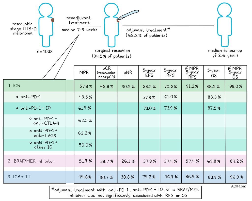
Neoadjuvant treatment with immunotherapy and/or targeted agents has become standard of care for resectable stage IIIB-D melanoma. However, the optimal treatment regimen and the consequences for long-term survival remain largely unknown. Long et al. analyzed a large dataset of patients treated with immune and/or targeted neoadjuvant treatments to answer some of these outstanding questions. Their results were recently published in Nature Medicine.
Data were collected from 1,038 patients with cutaneous, clinically detected, resectable, stage IIIB-IV melanoma who were treated with neoadjuvant systemic therapy across 26 international institutions from 2023 to 2024. Of these, 755 were participating in a clinical trial. Neoadjuvant treatment consisted of ICB in 70.8% of patients, BRAF/MEK inhibitors in 11.5% of patients, and a combination of ICB and a targeted agent (ICB + TT) in 17.7% of patients. The ICB cohort was treated with anti-PD-1 alone (23.3%) or with a combination of anti-PD-1 and another immune-oncology agent (PD-1 + IO), including anti-CTLA-4 (37.2%), anti-LAG3 (5.6%), and other IOs (4.3%). BRAF V600 mutations were present in 49.1% of patients.
Median follow-up from the first treatment was 2.6 years, and the median duration of neoadjuvant treatment was 7-9 weeks. Most patients underwent surgery (94.5%), and 66.2% received adjuvant treatment, including 47.9% receiving anti-PD-1, 4.7% receiving anti-PD-1 + IO, and 13.6% receiving BRAF/MEKis.
Pathological response was recorded in 959 patients. Major pathological responses (MPR), including pathological complete response [pCRs] or near-pCR), were observed in 57.8% of patients treated with ICB (pCR: 46.8%), 51.4% of those treated with BRAF/MEKis (pCR: 38.7%), and 44.6% of those treated with ICB + TT (pCR: 30.7%). Pathological non-response rates (pNR) were 30.5%, 26.1%, and 30.8%, respectively. MPR was significantly associated with treatment type after adjusting for confounders. Among patients receiving ICB, those treated with PD-1 + IO had higher MPR rates and lower pNR rates than those treated with anti-PD-1 alone. MPR was 62.5% for patients treated with anti-PD-1 plus anti-CTLA-4, 63.2% for anti-PD-1 and LAG3 inhibitors, and 50.0% for anti-PD-1 + other IO. In multivariate analyses, MPR was significantly associated with ICB treatment type.
Event-free survival (EFS) was defined as the time from the first dose of neoadjuvant therapy to the first occurrence of unresectable progression, recurrence, or death from any cause. The 5-year EFS was 68.5% for ICB (57.8% for anti-PD-1 alone, and 73.0% for combination ICB), 37.9% for BRAF/MEKis, and 74.2% for ICB + TT.
Recurrence-free survival (RFS) was defined as the time from surgery to first recurrence or death from any cause. The 5-year RFS rates were 70.6% for ICB (61.0% for anti-PD-1 alone, 73.9% for combination ICB), 37.4% for BRAF/MEKis, and 76.4% for ICB + TT. Patients with MPR had the highest 5-year RFS rates (91.2% for ICB, 57.4% for BRAF/MEKis, 89.6% for ICB + TT). Among patients treated with ICB, pCR was associated with a 5-year RFS of 92.7%, and pNR with 35.2%. The significantly lower RFS for neoadjuvant BRAF/MEKis suggest that ICB is the best neoadjuvant treatment option for patients with BRAF V600 tumors.
Overall survival (OS) was defined as the time from the first neoadjuvant dose to death from any cause. The 5-year OS rates were 86.5% for ICB (83.3% for anti-PD-1, 87.5% for combination ICB), 69.8% for BRAF/MEKis, and 83.9% for ICB + TT. Those with MPR had the highest OS rates (98.0%, 84.2%, and 96.9%, respectively); however, the interaction between neoadjuvant treatment and MPR was not significantly associated with OS. OS rates in ICB-treated patients were higher for those with pCR and lower for those with pNR. OS rates were equally high for patients achieving pCR after PD-1 alone or combination ICB. Overall, both pCR and near-pCR were associated with equal impact on survival, supporting MPR as an appropriate endpoint to monitor.
The researchers then evaluated the impact of follow-on adjuvant therapy in neoadjuvant ICB-treated patients using pathological response assessment. 52.6% of patients received adjuvant ICB (anti-PD-1 alone, n=309; combination ICB, n=49), 6.5% received BRAF/MEKis, and 41.0% received no adjuvant systemic therapy. In ICB-treated patients with MPR, RFS and OS rates were similar regardless of whether adjuvant ICB was administered. Adjuvant ICB also had no impact on RFS or OS among those with pPR (pathological partial response). Among those with pNR, adjuvant BRAF/MEKis and adjuvant ICB trended toward improved RFS and OS rates.
Of the patients treated with ICB, 38.5% had BRAF V600 mutations, 49.4% were BRAF wild-type, and 3.5% had non-V600 mutations. Patients with a V600 mutation who were treated with ICB had lower MPR rates than those with WT and non-V600 mutations. Patients with BRAF V600-mutant tumors treated with PD-1 + IO had higher MPR and pCR rates than those treated with anti-PD-1 alone. In multivariate analyses, the mutation was a poor predictive factor for pCR, but not MPR. Patients with the mutation had worse EFS and RFS, but their OS was similar to that of patients without the mutation.
There were 26 patients with acral melanoma who received ICB. Of these, 21 underwent pathological assessment and 8 had MPR. At 5 years, the EFS rate was 26.0%, RFS 28.5%, and OS 73.1%. Of the 6 patients who died, 5 were treated with anti-PD-1, and all had pNR or unresectable pre-operative progression.
The researchers then conducted a subanalysis of patients with metastases. Of 718 patients with stage III melanoma treated with ICB, 87.2% had nodal disease, 5.3% had in-transit metastases, and 7.5% had both nodal and in-transit metastases. The MPR rates were 58.2%, 66.7%, and 51.1%, and the 5-year EFS rates were 69.6%, 76.9%, and 63.8% for these three groups, respectively, with similar trends for RFS and OS. An additional 17 patients in the ICB cohort had stage IV disease, of whom 6 achieved pCR, 1 near-pCR, 2 pPR, and 8 pNR. At a median follow-up of 2.1 years, 82.4% of patients were alive. Of the small number of stage IV patients (17), 6 had a pCR, 5 of whom were still alive and recurrence-free at the last visit.
Comparing trial and non-trial patients showed similar rates of pCR and pNR, but non-trial patients had worse EFS and RFS, perhaps suggesting that the complex care needed for proper neoadjuvant treatment is a real-world variable to optimize. No survival differences were found between males and females.
This pooled analysis shows neoadjuvant combination ICB therapy results in high MPR rates and OS, suggesting pathological response is predictive for outcomes. Targeted therapy was least effective in the neoadjuvant setting, and ICB showed efficacy in patients with BRAF-mutant tumors. These data can help guide neoadjuvant treatment decisions for melanoma treatment.
Write-up by Maartje Wouters, image by Lauren Hitchings


