

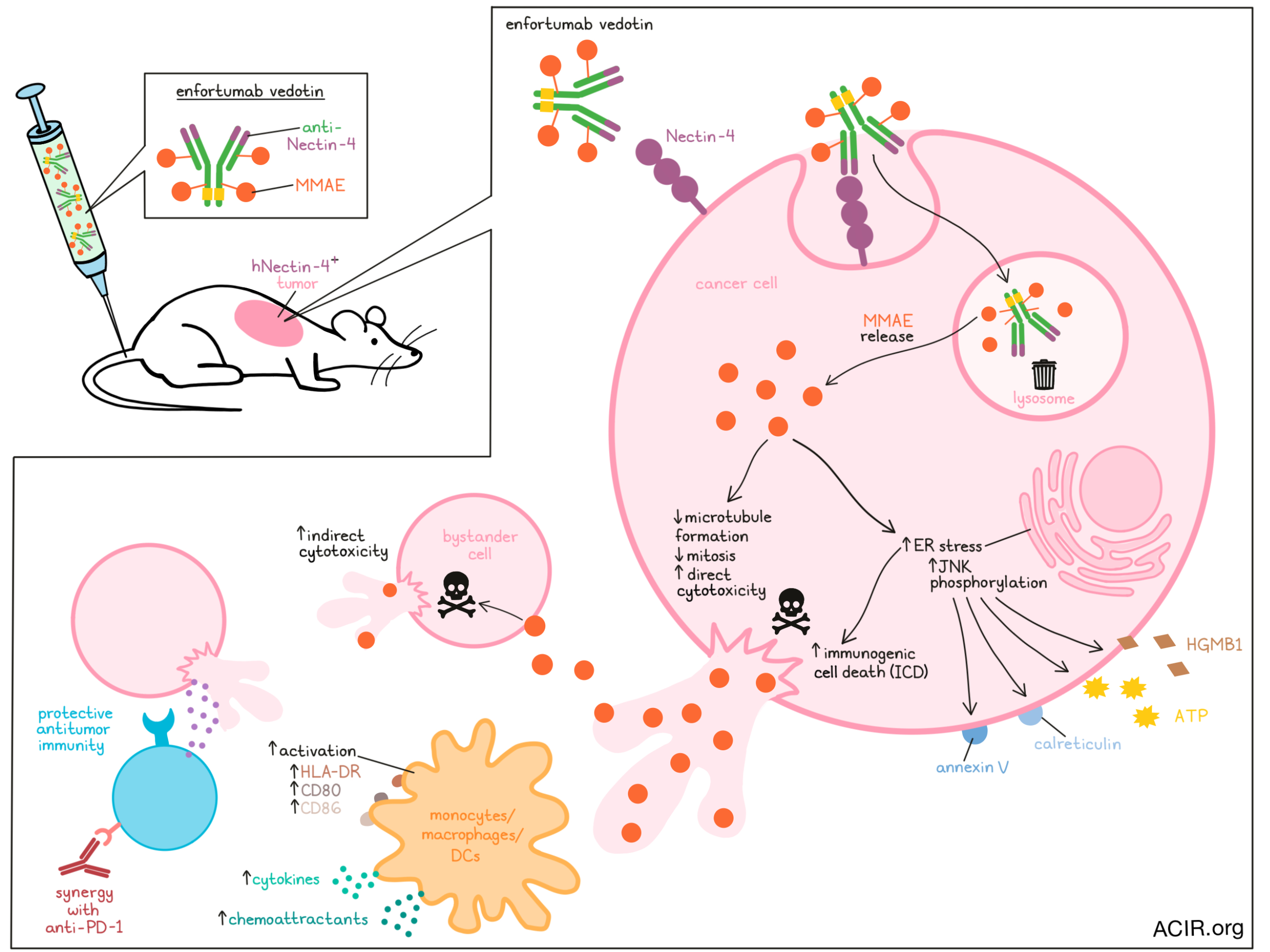
Enfortumab vedotin (marketed under the brand name Padcev) is an approved antibody–drug conjugate (ADC) that targets Nectin-4 on cancer cells and delivers a payload of microtubule-disrupting monomethyl auristatin E (MMAE) to tumor cells. In recent work, Olson and Liu et al. thoroughly investigated the mechanism of action of enfortumab vedotin, and identified direct, indirect, and immune-mediated antitumor activities. Their results were recently published in Cell Reports Medicine.
To begin, Olson and Liu et al. developed engineered human Nectin-4-expressing T24 cancer cells. Through indirect immunofluorescence, the researchers were able to observe hNectin-4-dependent internalization and trafficking of enfortumab vedotin to lysosomes within 2 hours, where enzymatic release of MMAE from the ADC occurs. The researchers then observed microtubule disruption and evidence of defective mitosis 48 hours after treatment, consistent with the direct cytotoxic effects of MMAE.
Next, the team investigated whether after its release from the ADC within Nectin-4+ cells, MMAE might have indirect cytotoxic effects on nearby hNectin-4- bystander cells. This was evaluated in a coculture model of hNectin-4+GFP- and hNectin-4-GFP+ M-UC-3 bladder cancer cells. After administration of enfortumab vedotin (compared to a non-binding IgG-MMAE control), the researchers observed dose-dependent cytotoxic effect and reduced viability of the hNectin-4-GFP+ population. Similar results were observed in a T24 bladder cancer cell coculture model.
In addition to direct and indirect cytotoxicity, MMAE can also induce ER stress resulting in immunogenic cell death (ICD), which could trigger an antitumor immune response. Investigating this possibility, Olson and Liu et al. evaluated ER stress by measuring JNK phosphorylation levels in T24-hNectin-4 cells treated in vitro with enfortumab vedotin, IgG-MMAE, or free MMAE. ER stress was evident starting at 24 hours, and by 48 hours, enfortumab vedotin induced the most ER stress (2.2-fold increase in phospho-JNK levels), followed by free MMAE (1.7-fold increase) and the non-binding control (1.5-fold increase). Other hallmarks of ICD were also observed, including increased extracellular release of ATP and HMGB1, and increased cell surface expression of annexin V and calreticulin. These effects were dependent on MMAE, and similar effects were observed in UM-UC-3-hNectin-4 cells.
Assessing potential immune responses to enfortumab vedotin-induced ICD, the researchers cocultured treated T24-hNectin-4 cells with purified CD14+ monocytes isolated from PBMCs. These monocytes showed increased expression of activation markers, including HLA-DR, CD86, and CD80. Similar but less pronounced effects were observed in monocytes cocultured with T24-hNectin-4 cells treated with free MMAE.
Next, the researchers evaluated whether this immune-activating effect would translate to enhanced antitumor immune responses in mice. In mice bearing xenograft Nectin-4-expressing human T24 urothelial tumors, the researchers found that a single i.p. injection of enfortumab vedotin reduced tumor growth. At 5 days post-injection, tumors showed increased F4/80+ macrophages and CD11c+ APCs compared to untreated controls, indicating recruitment of murine innate immune cells. Cytokines (IL-1α and CSF1) and chemoattractants (IP1β and MCP1) released by macrophages and DCs were also increased in enfortumab vedotin-treated tumors, as were interferon-inducible T cell chemoattractants (MIG and CXCL10). Further, treated tumors upregulated expression of genes associated with antigen processing and presentation, autophagy, type 1 interferon response, ER stress, myeloid DC activation and macrophage function. These results were consistent with the ICD induced by MMAE and with the induction of an inflammatory TME.
To untangle the cytotoxic effects of MMAE from the immune effects induced by ICD, the researchers generated an hNectin-4-expressing MB49 syngeneic mouse model. In vitro, MB49-hNectin-4 cells were treated with enfortumab vedotin in combination with the proton pump inhibitor valspodar, to prevent MMAE efflux and enrich for dying cells, and then sorted for dying cells with annexin V-conjugated microbeads. Vaccination with these cells protected mice from tumor engraftment in a majority of mice 14 days later, suggestive of immune-mediated protection. The same protection was not observed when mice were vaccinated with freeze/thaw tumor cells.
For certain clinical indications, enfortumab vedotin is approved for use in combination with pembrolizumab. To test the antitumor effects of this combination, the researchers utilized an MB49-hNectin-4 bladder carcinoma model and an EMT6-hNectin-4 breast cancer model. Following treatment, tumors in both models showed the greatest decrease in tumor volume and increase in macrophage infiltration after treatment with the combination of enfortumab vedotin and anti-PD-1. While 20% of the MB49 tumor-bearing mice treated with anti-mPD-1 alone rejected tumors, 35% rejected tumors when treated with the combination of anti-mPD-1 and enfortumab vedotin, resulting in durable complete tumor regression at primary tumor sites. When mice that had cleared their primary tumors were rechallenged with Nectin-4- parental tumor cells on the opposite flank, the majority (4 out of 5 in the anti-mPD-1 group and 7/9 in the combination group) rejected rechallenge, suggestive of durable antitumor immunity that was not necessarily specific to Nectin-4 target antigen expression, presumably due to epitope spreading.
Overall, Olson and Liu et al. shed light on the mechanism underlying the antitumor efficacy of enfortumab vedotin both alone and in combination with anti-PD-1. This clinically approved antibody–drug conjugate effectively targets Nectin-4 on cancer cells, which imports and transports the drug to the lysosome, where MMAE is released, disrupting microtubule formation and inducing ER stress and ICD in target cells. Once target cells die, MMAE is released, and can go on to kill nearby bystander cells, regardless of their expression of Nectin-4. Additionally, ICD stimulates a protective antitumor immune response against both Nectin-4+ and Nectin4- cancer cells. This enhanced understanding of this mechanism helps to validate the effects of enfortumab vedotin in clinical settings and may help to expand its application to additional settings moving forward.
Write-up and image by Lauren Hitchings


