

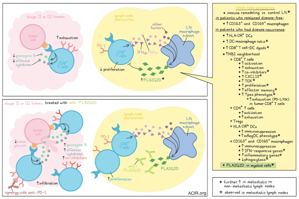
Tumor-draining lymph nodes (tdLNs) are critical sites for both antitumor immunity and tumor metastasis. To better understand what influences these events, van Krimpen and Huang et al. performed spatial proteogenomics studies on tdLNs from patients with melanoma. In addition to identifying a spatial neighborhood in the tdLN paracortex linked to poor prognosis, the researchers also identified a population of lymph node macrophages secreting PLA2G2D, which acted as an immune checkpoint that limited antitumor immunity and could be targeted for immunotherapy. These results were recently published in Nature.
Van Krimpen and Huang et al. began by collecting tdLNs from patients with resected stage II (non-metastatic to LNs) or III (metastatic to LNs) melanoma with known outcomes. Using an imaging mass cytometry (IMC) antibody panel, they evaluated regions of interest from various lymph nodes and identified individual immune cells based on marker expression. Compared to control LNs (from patients who died from acute vascular disease), tdLNs showed evidence of immune remodeling, even in the absence of metastasis, though the most substantial remodeling was evident in the paracortex of stage III tdLNs, with increased HLA-DR+ DCs in patients with recurrence and increased CD163+ and CD169+ macrophages in patients who remained disease-free. In metastatic tdLNs, increased DC:macrophage ratios were positively linked to disease recurrence.
Investigating interactions between cells in tdLNs, the researchers used nearest-neighbor analysis within the tdLN paracortex, and saw that several neighborhoods expanded or reduced in recurrent versus disease-free patients. While most cellular neighborhoods consisted of only one or two cell types, one neighborhood, defined as NB2, was characterized by increased CD4+ T cells, CD8+ cells, Tregs, HLA-DRhi DCs, and CD163+ and CD169+ macrophages, and was notably enriched in patients who experienced disease recurrence. T cells in NB2 showed increased markers of activation and exhaustion, while DCs and macrophages showed increased markers of immunosuppression. Further, DCs and CD8+ T cells were more likely to form dyads in recurrent patients, often with macrophages nearby.
Next, van Krimpen and Huang et al. performed a targeted spatial transcriptomics analysis to specifically evaluate the colocalization of DCs, CD8+ T cells, and CD68+ macrophages in paracortex regions. In samples from metastatic tdLNs, the researchers noted differentially expressed genes for each of the three cell types between patients with recurrence and those who remained disease-free. In patients with disease recurrence, DCs upregulated genes linked to a mature immunoregulatory (mRegDC) phenotype, while CD8+ T cells showed increased expression of genes or transcriptional signals for co-inhibitory molecules (CTLA-4, TGIT, PD-1), CXCL13, TOX, proliferation, and effector memory functions, in line with a progenitor exhausted T cell (Tpex) phenotype. Further, CXCL13 mRNA in paracortex CD8+ T cells correlated with the expression of PD-1 and TOX within tumors, suggesting that the Tpex phenotype in tdLNs predisposes T cells to exhaustion in tumors. In line with this, PD-1+CD8+ T cells more readily upregulated exhaustion-associated markers upon prolonged ex vivo stimulation. Macrophages from patients with recurrence upregulated IFN-responsive and immunosuppressive genes linked to an inflammatory phenotype, and reduced expression of genes associated with phagocytosis.
One gene that was notably upregulated in paracortex myeloid cells from patients with disease recurrence was for the secreted phospholipase PLA2G2D. In data from The Cancer Genome Atlas (TCGA), PLA2G2D was upregulated in melanoma samples, with the highest expression in metastatic LNs. Immunohistochemistry identified the highest expression of PLA2G2D in the interfollicular regions of the paracortex, particularly in patients with disease recurrence. Immunofluorescence staining in NSCLC samples also showed PLA2G2D+ macrophages and DCs in stage II and stage III NSCLC tdLNs, with much lower levels in primary tumors or LN metastases. In tdLNs, PLA2G2D+ myeloid cells were also frequently located near PD-1+CD8+ T cells.
Investigating the role of PLA2G2D, the researchers evaluated tumor growth in wild-type and Pla2g2d-deficient (Pla2g2d−/−) mice, and found that several different tumor models showed reduced growth in Pla2g2d−/− mice, which could be reversed by administering PLA2G2D–Fc protein. In vitro, treating CD4+ or CD8+ T cells from healthy donor peripheral blood with PLA2G2D–Fc reduced TCR stimulation-induced proliferation, effector cytokine production, and granzyme B release. These effects were independent of Fc function and the enzymatic activity of PLA2G2D, suggesting that signaling may occur through an unknown receptor on T cells.
In an effort to improve antitumor immune responses, van Krimpen and Huang et al. generated both mouse and human antibodies that blocked PLA2G2D. These antibodies reduced tumor growth in murine and humanized mouse tumor models, respectively, including metastatic tumor models.
To determine which cells produce PLA2G2D in vivo, the team generated bone marrow chimeras and showed that cancer progression was reduced in wild-type mice with Pla2g2d−/− bone marrow, but not Pla2g2d−/− mice with wild-type bone marrow. Single-cell transcriptomics identified strong Pla2g2d in a subset of macrophages that was highly abundant in tdLNs, but undetectable in primary tumors. Further, Pla2g2d served as an identity-defining gene for this subset, which resembled macrophages identified in tdLNs of patients with disease recurrence, as well as previously identified T cell zone macrophages. The subset was also confirmed in scRNAseq data from patients with other tumor types.
To identify factors that induce PLA2G2D, the researchers treated monocyte-derived macrophages with the supernatant of T cell cultures, or with IFNγ and TNF, both of which effectively induced PLA2G2D expression. IFNγ and TNF also correlated with PLA2G2D in several tumor types in TCGA, suggesting that T cell-derived inflammatory cytokines induce myeloid PLA2G2D. However, in mouse tumor models, IFNγ neutralization did not reduce lymph node macrophages or Pla2g2d expression, suggesting that other factors also contribute to the induction of PLA2G2D.
When the researchers treated tumor-bearing mice with anti-PLA2G2D antibodies, they noted increased expression of proliferation markers and co-inhibitory molecules on CD8+ T cells, including tumor-specific and memory cells. Anti-PLA2G2D also enhanced CD8+ T cell infiltration into primary tumors, and in depletion studies, CD8+ T cells were shown to be required for treatment benefit.
The treatment benefits of anti-PLA2G2D were not durable, and later tumor progression coincided with high levels of PD-1+CD8+ T cells in the blood, tdLN, and tumor. When anti-PLA2G2D (or genetic ablation of PLA2G2D) was combined with anti-PD-1, tumor growth was further reduced compared to either monotherapy in both murine and humanized models. Combination therapy also increased tumor-specific IL-2-producing CD8+ T cell infiltration. These results suggest that PLA2G2D and PD-1 are non-redundant checkpoints, and that there were benefits to targeting both in combination.
Overall, these results suggest that PLA2G2D, secreted primarily by a subset of lymph node macrophages, acts as a targetable immune checkpoint by suppressing early CD8+ T cell functionality. Blocking PLA2G2D reduced this immunosuppression and enhanced antitumor immunity, resulting in reduced tumor growth, which could be further reduced in combination with anti-PD-1. PLA2G2D was associated with poor prognosis in patient data, further indicating the potential for future clinical applications.
Write-up and image by Lauren Hitchings
Meet the researcher
This week, co-first author Anneloes van Krimpen answered our questions.

What was the most surprising finding of this study for you?
Previous research hinted that this specific region of the tumor-draining lymph nodes (tdLN) may be altered in cancer. However, we didn’t expect the extent to which these regions were remodeled in association with disease recurrence. When we further characterized cells within this tissue location, PLA2G2D was a standout hit. Although our initial cohort was small, we found this same population and predominance of PLA2G2D+ cells in other cancers, suggesting a cancer-overarching mechanism. That Apeximmune Therapeutics had developed an antibody to target it, parallel and independent to our own findings, was the cherry on top.
What is the outlook?
In future research, we hope to better understand what conditions create an environment that potentiates immunosuppressive cells, such as PLA2G2D+ myeloid cells, within the tdLN, and how this is tied to the primary tumor. Furthermore, in murine models across multiple cancer types, targeting PLA2G2D significantly reduced tumor growth, especially when combined with anti-PD1 therapy. Our data also suggest that this approach may help overcome resistance to anti‑PD‑1, potentially enhancing the efficacy of current immunotherapies. Our collaborators at Apeximmune Therapeutics are now working to move this approach toward clinical trials.
Who or what has been a major source of inspiration or motivation for you throughout your career?
My biggest inspiration and motivation have always been people. It’s the patients who participate in research, my coworkers, who supported me and with whom I’ve formed close friendships, my supervisors who believed in me, and my family who cheered me on. I’ve worked on this project for six years, and it’s easy to lose sight of why exactly you’re doing this when things get hard. Seeing the reception of our research, especially from patients with cancer or those who have lost their loved ones to it, made me appreciate what a privilege it is to do this kind of job.


