

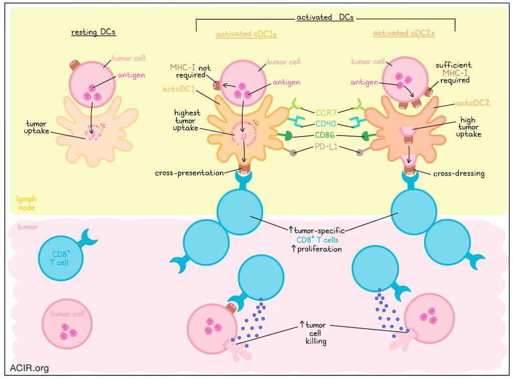
A subset of cDCs within tumors expresses T cell-stimulating and immune-regulatory molecules. The exact role of these activated DCs (actDCs) in antitumor immunity remains largely unknown, and given their scarcity, they are a challenge to study. Koufaki et al. developed murine models to label and ablate CCR7+ DCs to study their role in antitumor immunity. Their results were recently published in Immunity.
Transcriptomic profiling of DCs from 5555 BrafV600E-driven melanoma or CT26 colorectal carcinoma revealed four clusters: two clusters comprising conventional DCs (cDC1s and cDC2s), plasmacytoid DCs (pDCs), and a cluster with high expression of genes such as Ccr7, Cd80, and Cd40. Furthermore, expression of transcription factors in this latter cluster indicated DC activation. The CCR7+ cDCs exhibited lineage-defining markers of cDC1 and cDC2 at lower levels, while showing higher expression of markers such as CD40, CD86, and PD-L1. This suggested that the population represented an activation state shared by cDC1s and cDC2s (actDCs).
Given their scarcity, the researchers developed a genetically engineered mouse line to selectively label, track, and isolate actDCs. Because Ccr7 was detected in actDCs, but not in cDCs, the researchers engineered a mouse line in which actDCs expressed the fluorescent protein mScarlet-I (Scarlet) to enable fluorescent labeling of actDCs. Crossing this line with the CD11c-Cre driver generated actDC-Sca animals. Four syngeneic cancer models were implanted in these mice, and Scarlet expression was detected in 20-40% of tumoral cDC1s and cDC2s.
The researchers then investigated whether tumoral actDCs could also be obtained in conventional FLT3L-BMDC cultures. Bone marrow from actDC-Sca mice underwent a standard cDC differentiation protocol, yielding a Scarlet+ population. Adding live 5555 melanoma cells to the differentiated DCs in vitro increased Scarlet expression in both cDC1s and cDC2s. scRNAseq of these in vitro-generated actDCs confirmed their similarity to tumor-infiltrating actDCs, revealing a cluster of Ccr7-expressing cells lacking lineage-defining markers and enriched for the in vivo actDC signature.
For functional characterization of actDCs, OVA-expressing 5555 cells were added to FLT3L-BMDC cultures from actDC-Sca mice, either with actDCs or resting DCs. The next day, their ability to present cancer-associated antigens and stimulate proliferation of OVA-specific T cells was assessed. Only actDCs robustly induced OT-I proliferation, with actcDC1s and actcDC2s showing similar effects. This was also true when DCs were obtained from tumor-draining lymph nodes (TDLN) of actDC-Sca mice with 5555OVA tumors. Using ZsGreen-expressing 5555 cells, imaging analysis showed that ZsGreen could be acquired and localized intracellularly in all DCs, but mainly in the ActDC1s. However, uptake of tumor cell-derived material was not a prerequisite for actDC differentiation, and the difference in priming between actDCs and resting DCs could not be explained by differences in antigen acquisition.
Since cDC2s could prime CD8+ T cells, the researchers investigated whether cross-dressing, rather than cross-presentation, was responsible for these effects. β2m was knocked out in 5555OVA cells to generate MHC-I-deficient tumor cells that could not cross-dress cDCs. These cells were inoculated into actDC-Sca mice, and sorted DCs from the TDLNs were used for ex vivo OT-I cultures. Only the actDC1s induced OT-I proliferation in this setting, suggesting that actDC2s rely on cross-dressing. To assess the in vivo role of cross-dressing in priming, endogenous tumor-specific CD8+ T cell responses were assessed in TDLNs of WT mice bearing MHC-I-sufficient and -deficient 5555OVA tumors. The MHC-I-sufficient tumors were rejected, whereas the deficient ones progressed, with lower frequencies of tumor-specific CD8+ T cells, suggesting that cross-dressing during priming was more limited.
To determine if cross-dressing becomes more important when cDC1-mediated cross-presentation is limited, cDC1-deficient and cross-presentation-deficient mice were generated. Both mouse models developed progressive 5555OVA tumors. When MHC-I-sufficient or -deficient 5555OVA cells were injected, followed by naive OT-I transfer, OT-I activation was reduced in the deficient mice, but was similar in WT mice. This suggested that cross-dressing played a role in T cell priming when cDC1-driven cross-presentation was limited.
To assess differences in cross-dressing in other tumor types, the researchers generated OVA-expressing B16 melanoma cells and cocultured them with BMDCs. DC populations were then sorted to determine their impact on OT-I proliferation. Only actDC1s stimulated OT-I cells in this setting. Both 5555OVA and B16OVA cell lines have similar MHC-I expression; however, when the B16 line was pre-treated with IFNγ to increase MHC-I, actcDC2s were able to induce OT-I proliferation.
Koufaki et al. then investigated the role of actDCs in antitumor immunity and generated a conditional strain in which ablation of CCR7+ DCs occurs upon diphtheria toxin (DT) treatment (ActDC-DTR). This strain was used to test the role of actDCs in 5555OVA antitumor immunity. Tumors regressed in untreated mice, but progressed after DT treatment. DT reduced the number of circulating tumor-specific CD8+ T cells and the number of intratumoral T cells.
To assess the role of actDCs in tumor-specific CD8+ T cell priming, naive OT-I cells were transferred into 5555OVA-bearing actDC-DTR mice, and their activation was assessed 16 hours later. In control mice, OT-I cells showed robust activation, whereas activation was minimal after actDC depletion, suggesting that early priming depends on the actDC state.
Finally, the researchers assessed the contribution of actDCs to immunotherapy. First, immune checkpoint blockade (ICB) was assessed by inoculating actDC-DTR mice with 5555 cells and treating them with dual CTLA-4 and PD-1 ICB, which eradicated tumors. When actDCs were depleted, 40% of mice exhibited progressive tumor growth and had fewer intratumoral CD4+ and CD8+ T cells. Second, adoptive cell transfer (ACT) was tested using actDC-DTR strains on a Rag1-/- background, which removes endogenous T and B cells. 5555OVA inoculation induced aggressive tumors, and a single infusion of in vitro-expanded OT-I cells caused tumor shrinkage. ActDC depletion reduced ACT efficacy, with two-thirds of mice showing minimal or no benefit, and their OT-I cells expressing dysfunction markers.
Overall, these data highlight the importance of CCR7+ actDCs in antitumor immunity, with both actDC1s and actDC2s contributing to T cell-mediated antitumor responses. Furthermore, because these cells were shown to be important determinants of immunotherapy efficacy, they may serve as potential new therapeutic targets.
Write-up by Maartje Wouters, image by Lauren Hitchings
Meet the researcher
This week, first author Maria Koufaki answered our questions.

What was the most surprising finding of this study for you?
One of the biggest surprises was discovering just how essential the CCR7⁺ activated state of dendritic cells is for antitumor immunity. We initially expected activated dendritic cells (actDCs) to contribute to T cell priming, but the extent of their non-redundant role was striking. Despite representing only a small fraction of dendritic cells within tumours, removing actDCs almost abolished spontaneous antitumor immunity and severely impaired responses to immune checkpoint blockade and adoptive T cell therapy. These findings reveal the activation state of dendritic cells as a pivotal, non-redundant component of effective antitumor immunity.
What is the outlook?
A major next step is to understand what drives dendritic cells into this activated state, and whether we can therapeutically manipulate it. Much of cancer immunotherapy has focused on boosting T cell responses, but our findings suggest that generating or sustaining the activated state of dendritic cells may be just as important for effective antitumor immunity. We hope these new mouse models will become a useful resource for the wider immunology community to explore this biology, not only in cancer, but also in infection and autoimmune disease.
What was the coolest thing you’ve learned (about) recently outside of work?
I recently started playing tennis after years of playing volleyball (I thought it would help more than it did!). It has been a good reminder that starting something new is both rewarding and humbling. Progress feels slow until you hit, every so often, a great rally that reminds you why you started in the first place. Not that different from research.


