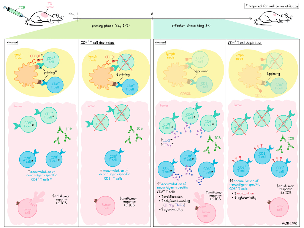
While CD4+ T cells are crucial for CD8+ T cell priming in ICB-induced antitumor responses, their helper role at the effector phase remains largely unknown. Ameh et al. used murine MHC-II- sarcoma models to study CD4+ T cell help during ICB treatment. Their results were recently published in Cancer Immunology Research.
The researchers had previously shown through CD4+ T cell depletion that CD4+ T cells are essential for ICB-induced rejection of MCA-induced T3, F244, and 1956 sarcomas. To understand their role separately in the priming and effector phases, the team first examined T cell accumulation in untreated T3 tumor-bearing mice. Neoantigen-specific CD4+ and CD8+ T cells accumulated in tumors by day 7 post-transplantation, with limited further increase.
Based on these results, day 8 was chosen as the time point to distinguish between early (priming phase) and post-priming (effector phase) events. To establish the role of CD4+ T cells during the priming phase, a CD4-depleting antibody was administered 1 day before tumor inoculation and continued thereafter. This reduced accumulation of mLama4-specific CD8+ tumor-infiltrating lymphocytes (TILs), and no tumors responded to ICB, suggesting impaired CD8+ T cell priming or recruitment to the tumor.
To assess the role of CD4+ T cells during the effector phase, anti-CD4 was initiated on day 8 after tumor inoculation, depleting CD4+ T cells within 24 hours. On day 9, numbers of mLama4-specific CD8+ TILs were similar in depleted and control mice, suggesting priming had occurred. Yet, despite priming, ICB treatment did not cause tumor rejection, highlighting that CD4+ T cells are vital for both priming and the effector phase of antitumor CD8+ T responses during ICB.
The researchers then determined whether CD4+ T cells were required at the tumor site to support CD8+ T cell function after priming. Tumor-bearing mice received FTY720 to block lymphocyte egress from lymphoid tissues. When given during priming, no mice rejected the tumor in response to ICB. When given during the effector phase, starting on day 8 when T cells had accumulated at the tumor, ICB induced rejection in normal mice. Depleting CD4+ or CD8+ T cells during this phase prevented ICB-induced tumor rejection, indicating that once T cells are primed and reach the tumor, ongoing peripheral priming is not required.
Ameh et al. hypothesized that CD4+ T cells in the tumor mediate the reinvigoration of tumor-specific CD8+ T cells during the effector phase. To test this, the numbers and phenotypes of T3 neoantigen-specific CD8+ T cells were assessed in mice treated with dual ICB with or without late CD4 depletion. ICB treatment increased total and mLama4-specific CD8+ T cells and Ki67+CD8+ T cells, an effect that was independent of CD4+ T cells. However, mLama4-specific CD8+ T cells in CD4-depleted mice had higher levels of exhaustion markers than non-depleted mice. The exhaustion marker expression was at similar levels as found in untreated mice, reflective of tumor progression.
Dual ICB increased the frequency of polyfunctional (IFNγ+TNFα+) mLama4-specific CD8+ T cells, but these cells were absent in mice receiving a late CD4 depletion. Similarly, the ex vivo cytotoxic potential of these CD8+ T cells was impaired when obtained from ICB-treated mice depleted of CD4 at the effector stage. These data suggest that CD4+ T cells are required to reinvigorate CD8+ T cell effector function during ICB treatment.
To determine whether MHC-II presentation is essential in the effector phase, the researchers developed a TCR-mimic antibody targeting the mutant Itgb1:I-Ab complex that blocks CD4+ T cell activation at the level of peptide presentation on MHC-II. To do this, the team generated a panel of human monoclonal antibodies (mAbs) targeting the dominant T3 Itgb1 MHC-II neoantigen bound to the I-Ab molecule. The human mAb with the greatest inhibitory activity was selected and re-engineered with a mouse IgG1 sequence for the murine experiments. The mAb, m1G10, blocked mltgb1-specific hybridoma activation by antigen-presenting cells (APCs) and inhibited IFNγ production by polyclonal CD4+ T cells isolated from mice vaccinated with mltgb1 synthetic long peptide, while it did not impact the CD4+ T cells from mice vaccinated with an irrelevant neoantigen.
Antibody staining of T3 tumor tissue showed that macrophages, cDC2s, and cDC1s present this complex to CD4+ T cells. To determine whether MHC-II neoantigen presentation impairs CD4+ T cell function at the effector phase, a simplified system based on the T3 model in RAG2-/- mice was used, as T3 tumors have a high mutational load and may have multiple MHC-II epitopes. In this model, tumor rejection is driven by adoptive transfer of CD4+ and CD8+ T cells primed by vaccination against the two dominant MHC-I-restricted neoantigens of T3 and the dominant T3 MHC-II neoantigen. However, when m1G10 was administered, or CD4+ or CD8+ T cells were depleted, tumors were not rejected.
The researchers proposed two hypotheses regarding the function of CD4+ T cells in the effector phase: I) licensing DCs via cell-to-cell contact mediated by CD40/CD40L interactions, or II) producing cytokines such as IL-2 and IFNγ. CD40L blockade during the priming phase eliminated the antitumor effects of ICB, whereas late blockade during the effector phase had no impact. Blockade during priming reduced the total number of mLama4-specific CD8+ TILs and decreased IFNγ-producing mLama4-specific CD8+ T cells in the spleen and tumor-draining lymph nodes. T3 tumor-bearing mice treated with ICB and blocking antibodies for IFNγ and IL-2 did not reject tumors. Therefore, CD40/CD40L signaling is essential for priming, and IL-2 and IFNγ are essential for ICB-induced antitumor CD8+ T cell responses during the effector phase.
Together, these data shine a light on the function of CD4+ T cells in ICB-induced antitumor immunity, suggesting CD4+ T cell help is required during CD8+ T cell priming and during the effector phase in the tumor. These data may help in the design of future cancer immunotherapy strategies, particularly combination strategies.
Write-up by Maartje Wouters, image by Lauren Hitchings
Meet the researcher
This week, first author Samuel Ameh answered our questions.
What was the most surprising finding of this study for you?
The most surprising finding was the extent to which CD8⁺ T cells depended on CD4⁺ -cell help during immune checkpoint therapy (ICT). We initially expected that removing CD4⁺ T cell help during the effector phase would substantially reduce the expansion or accumulation of tumor-specific CD8⁺ TILs. Instead, CD8⁺ TILs still expanded and accumulated within the tumor, but they lacked the functional quality required for effective tumor elimination. This showed that CD4⁺ T cells were required for the qualitative, but not quantitative, enhancement of CD8⁺ TILs by ICT.
What is the outlook?
Our findings emphasize the importance of cooperation between CD4⁺ and CD8⁺ TILs for antitumor immunity during ICT. CD8⁺ TILs require CD4⁺ T cell help to function optimally not only during priming, but also within tumors during the effector phase, even in the presence of ICT. An important next step is to define how CD4⁺ T cells directly and indirectly support CD8⁺ TIL function and determine whether these pathways can be therapeutically enhanced. This could guide strategies to improve ICT in patients whose tumors contain CD8⁺ TILs, but fail to respond, while supporting treatments that preserve CD4⁺ and CD8⁺ T cell cooperation.
What was the coolest thing you have learned recently outside of work?
Through Jeopardy!, I recently learned the meaning behind the traditional red-and-white barber pole. The red is associated with blood, while the white represents the bandages used during bloodletting procedures. The pole itself is thought to symbolize the staff that patients gripped to make their veins more prominent. The symbol dates back to a period when barbers performed not only haircuts, but also surgical and medical procedures, a rather fascinating union of vocations. I had seen barber poles countless times in the USA without really considering their origin, so learning that such an ordinary symbol preserves this history was especially memorable.