

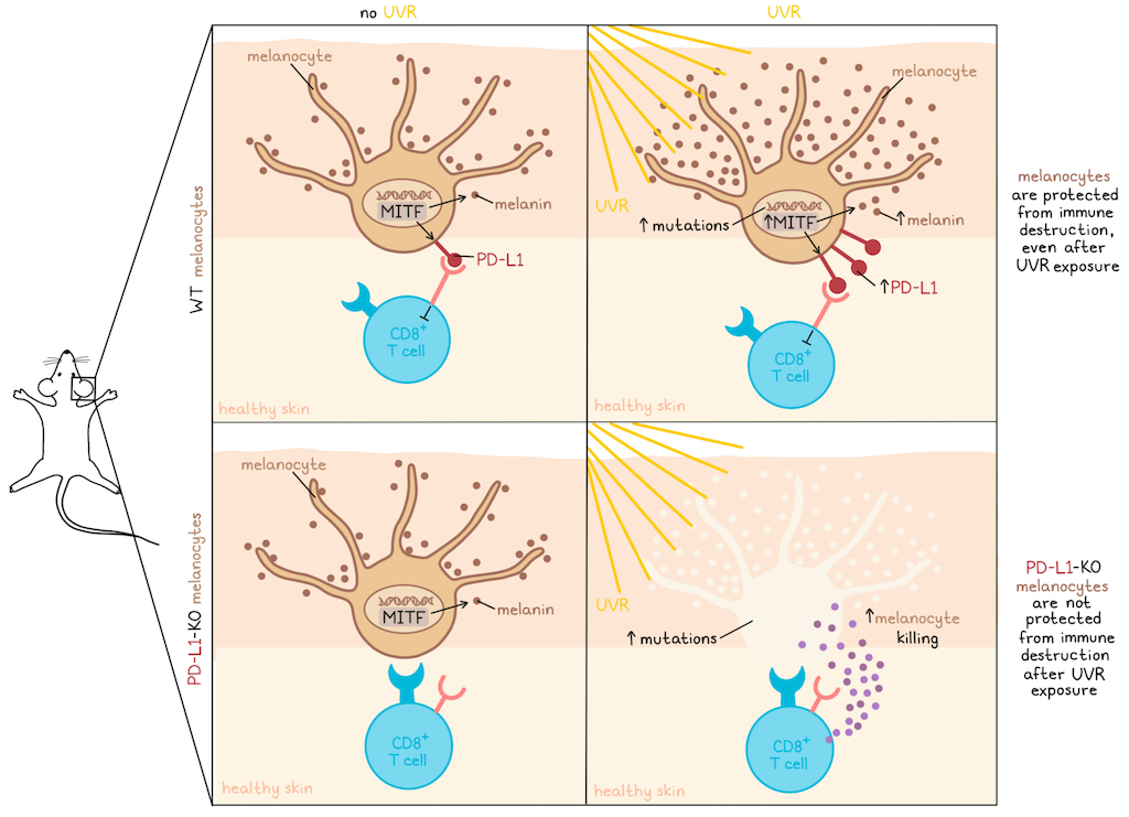
Melanocytes in human skin are regularly exposed to ultraviolet radiation (UVR) from the sun, and can accumulate substantial DNA damage without being eliminated by the immune system. In recent work published in Immunity, Lo et al. investigated how these cells evade immune elimination, and found that the transcriptional regulator microphthalmia-associated transcription factor (MITF), which is crucial to the melanocyte identity, induces PD-L1 at baseline and further upregulates it upon UV exposure, protecting melanocytes from CD8+ T cell-mediated destruction.
Following prior evidence that MITF expression correlated with PD-L1 expression in patient melanomas, Lo et al. began by evaluating the chromatin landscape in primary human melanocytes upon silencing of MITF. Among all genome-wide MITF binding sites, H3K27ac signals at the site of the PD-L1 gene were among the most diminished upon MITF silencing. More specifically, there was a significant H3K27ac enrichment peak shortly upstream of the PD-L1 transcription start site that contained 3 consensus MITF binding sites (E-boxes), and was lost upon MITF silencing, identifying a putative enhancer. Further, MITF silencing reduced PD-L1 expression at the RNA and protein levels, while overexpression of MITF upregulated PD-L1 expression. MITF protein occupancy was also confirmed at the site of the putative enhancer. Similar results were identified in other datasets and in experiments using CUT&RUN profiling and a luciferase reporter model in which luciferase activity was repressed by MITF silencing and stimulated by MITF overexpression in melanoma cells. These effects were lost upon mutation of either of the the two E-boxes located within the identified MITF binding site peak, but not of the E-box outside the peak. Together, these data suggested that MITF directly mediated PD-L1 transcription by engaging with E-box elements in an upstream enhancer.
Next, the researchers investigated the role of MITF regulation of PD-L1 at baseline in healthy skin by evaluating mice lacking MITF, and thus, melanocytes. In humans, melanocytes are located below the epidermal cells in the majority of skin, but in mice, they are primarily found in hair follicles over most of the fur-covered body. Thus, the researchers looked at the ears, where there is no fur, and melanocytes are present below the epidermis, as they are in humans. Here, the researchers found that skin from mice lacking MITF/melanocytes showed lower levels of PD-L1, consistent with baseline PD-L1 levels in other skin cell types.
Investigating whether MITF may upregulate PD-L1 in response to UVR, as it does with melanin in the tanning response, the researchers evaluated human skin explants under UVB conditions (which represent the majority of UVR) consistent with strong daily sun exposure, and found that both MITF and PD-L1 expression increased. Increased PD-L1 expression following UV exposure was also observed in mouse models, and the effect was largely abrogated in mice lacking MITF/melanocytes.
Further exploring the induction of MITF-induced PD-L1 upregulation, Lo et al. noted that UVR-mediated induction of the MITF tanning pathway in melanocytes is mediated indirectly via p53 activation in other skin cells. Following this logic, the researchers found that conditioned medium from UVR-exposed keratinocytes could increase MITF and PD-L1 expression in melanocytes. Additionally, as type I IFNs and IFNγ can induce PD-L1 in melanomas and other cancers, the researchers evaluated IFNγ/IFNAR-deficient mice, but found no differences in the response to UVR compared to wild-type mice. IRF1, which induces PD-L1 transcription in response to IFNγ signaling, was also not required for MITF-driven PD-L1 upregulation in melanocytes, suggesting that the mechanism is not IFN-dependent.
Lo et al. then turned to patient data to determine whether there might be a link between MITF and PD-L1 expression in melanoma. While the correlation between IFNγ and PD-L1 dominated the data for melanomas with high IFNγ signaling, in melanomas with lower CD8+ T cell infiltration and cytolytic scores, the researchers were able to identify a significant correlation between MITF and PD-L1 mRNA expression. These results suggested that while there may be a role for MITF-induced PD-L1 in more immune-cold melanomas, its role may be more pertinent as a tolerogenic mechanism at baseline for protecting highly UV-mutated melanocytes from immune-mediated destruction.
Next, the researchers studied the effects of PD-L1- melanocytes using humanized mouse models with PD-L1 conditionally knocked out in melanocytes. Mice were exposed to periodic UVB radiation consistent with strong sun exposure over the course of 16 weeks. After cessation of UVB treatments (which have a known anti-inflammatory effect), mice lacking PD-L1 in melanocytes showed significantly less ear pigmentation than controls, which could be attributed to an observed increase in CD8+ T cells and a reduction in melanocytes in the epidermis. These results suggested that in the absence of PD-L1, UV-exposed melanocytes were more susceptible to immune-mediated clearance. No differences were observed in the absence of UVR, suggesting that an environmental stressor was required for the loss of PD-L1-mediated protection to become apparent.
Given that the pattern of immune-mediated depigmentation observed with PD-L1 loss was reminiscent of vitiligo, the researchers established a PD-L1-deficient adoptive transfer model of vitiligo. Compared to wild-type mice, mice lacking PD-L1 showed signs of depigmentation earlier after transfer of melanocyte-specific T cells. In vitro T cell killing assays showed higher IFNγ release and target cell killing against PD-L1-deficient melanocytes compared to those with intact PD-L1. Similar results were observed in cocultures of human iPSC-derived melanocytes and CD8+ T cells, further validating the protective effects of PD-L1 against immunity.
Finally, the researchers utilized human iPSC-derived melanocytes with or without PD-L1 expression, cocultured them with primary human dermal and epidermal cells, and engrafted the mixture onto the skin of immunodeficient mice. Upon adoptive transfer of gp100-specific human T cells into these models, PD-L1-deficient melanocytes were more readily eliminated than those with typical PD-L1 expression by one month after transfer. This effect was maintained after two months, suggesting long-term melanocyte depletion.
Overall, Lo et al. showed that in melanocytes, MITF mediated upregulation of PD-L1 by binding to an enhancer, inducing high baseline PD-L1 that was further upregulated upon exposure to UVR. This mechanism ultimately had a tolerogenic effect, protecting melanocytes from immune-mediated elimination, even after the accumulation of UVR-induced mutations. While this mechanism did not appear to play a major role in immune-hot melanomas with high IFNγ expression, it did show an effect in less infiltrated melanomas, and could contribute to their early development.
Write-up and image by Lauren Hitchings
Meet the researcher
This week, lead author David Fisher answered our questions.
What was the most surprising finding of this study for you?
The most surprising finding to me was that a gene as ubiquitously expressed at PD-L1 could be transcriptionally regulated by a lineage-restricted transcription factor. Because of this surprising feature, we at first did not quite believe the data made sense. Then, we realized this may be a mechanism to offer lineage-selective immune tolerance, since melanocytes constantly accumulate UV mutations that could target them for immune destruction. Therefore, a lineage-specific mechanism of tolerance suddenly felt plausible.
What is the outlook?
We can imagine that similar lineage-specific mechanisms of immune tolerance exist in other tissues – often we know this occurs through recruitment of immune modulating cell types (i.e., immunosuppressive cells). We wonder whether this mechanism may play an interesting role in autoimmune conditions involving melanocytes (i.e., vitiligo).
What was the coolest thing you’ve learned (about) recently outside of work?
One of the coolest things I recently learned about was how one of our sons (who has a PhD in Organic Chemistry) discovered a deep love for Sauna/Cold-Plunge and decided to open a spa for this in his hometown of San Diego.


