While accumulating evidence suggests an important role for CD4+ T cells in antitumor immunity, the specific subpopulations responsible remain to be defined. Agesta, Ferrand, et al. investigated the function of the transcription factor Eomes in CD4+ T cell antitumor responses, and identified Eomes-responsive antitumor Th populations, with significant analogies to similar Eomes-responsive CD8+ populations. Their findings were recently published in Immunity.
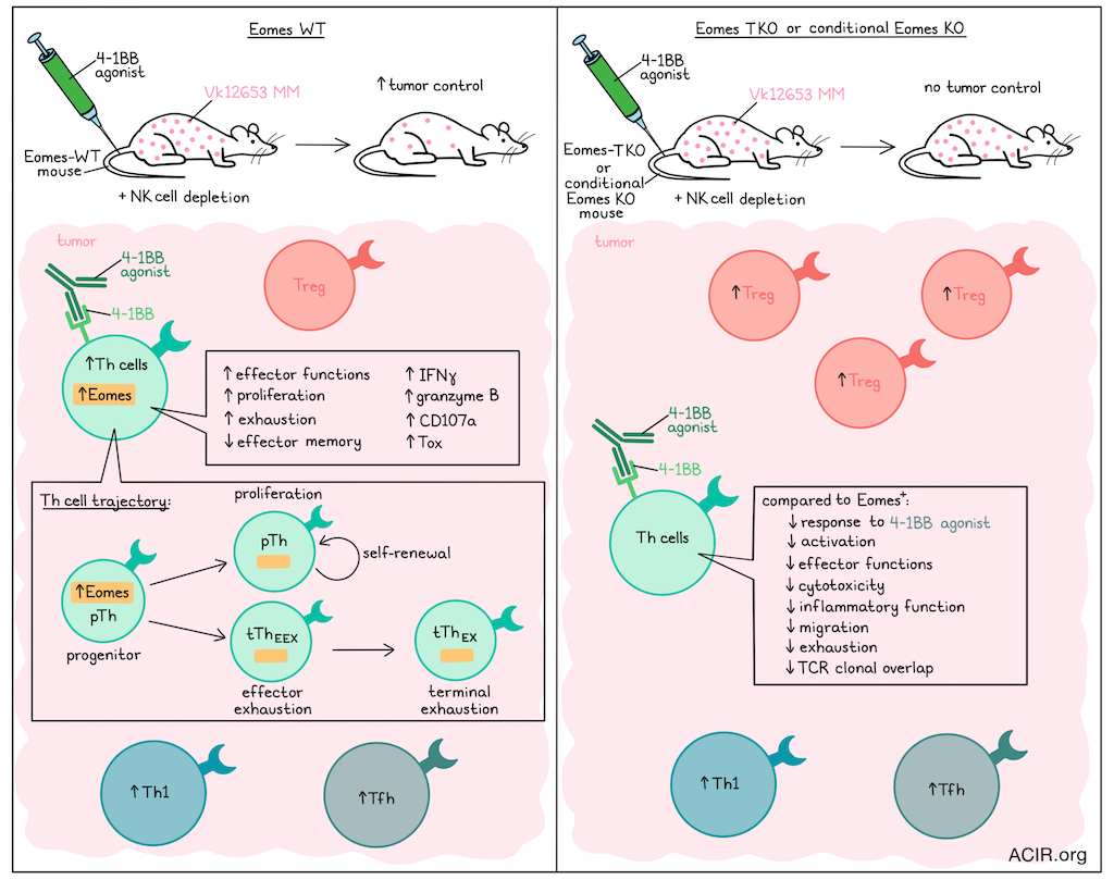
The researchers began by studying the role of Eomes in CD4+ T cells in tumor immunity, using conditional CD4+ T cell Eomes-deficient mice (Eomes-TKO) and WT mice (Eomes-TWT). Mice underwent NK cell depletion (to allow tumor cell engraftment), were inoculated with Vk12653 multiple myeloma cells, and were treated with a 4-1BB agonist. 4-1BB stimulation induced tumor regression and lowered serum gamma-globulin levels in the Eomes-TWT mice only. Eomes-TWT mice had increased levels of Th cells in the tumor, while Tregs were not affected. In Eomes-TKO mice, on the other hand, Th cells were not impacted, but there was an increase in Tregs in response to 4-1BB. Eomes-deficient Th cells from 4-1BB-treated mice had reduced activation and effector functioning.
4-1BB increased Eomes expression in Th cells. To determine whether this upregulation impacted tumor control independent of CD8+ T cell activity, RagγcKO mice were reconstituted with CD4+ T cells from the Eomes-TWT or Eomes-TKO mice, engrafted with Vk12653 cells, and 4-1BB was stimulated. Those mice reconstituted with Eomes-TKO cells showed no tumor regression, while Eomes-TWT CD4+ Th cells infiltrated the tumor, induced tumor regression, elevated Eomes levels, and exhibited enhanced effector function.
To determine how Eomes impacts Th cell fate, Eomes-GFP and Eomes-TKO mice were crossed with Foxp3-Thy1.1 reporter mice, to allow for exclusion of Tregs in the analyses. The resulting mice were engrafted with Vk12653 cells and treated with 4-1BB. Among TCR+CD4+Thy1.1-CD44hi T cells, effector, memory, and GFP+ clusters expanded upon 4-1BB treatment. GFP+ Th cells were highly proliferative and accumulated in the bone marrow (BM) and spleen. A Th subset analysis in the BM identified Tfh and Th1 cells distinct from the GFP+ cluster, which expanded over time in response to 4-1BB and were not affected by Eomes deletion.
The GFP+ Th cells expressed higher levels of exhaustion markers than effector or memory Th cells. The exhaustion features were already detectable in GFP+ Th cells from control mice not receiving 4-1BB, and these features increased over time following 4-1BB stimulation.
Bulk RNAseq was performed to determine the transcriptional program associated with Eomes expression in Th cells. Eomes-deficient or GFP- Th cells were minimally impacted by 4-1BB stimulation. GFP+ Th cells, on the other hand, underwent transcriptomic reprogramming, defining a unique Eomes-dependent signature. Effector/memory-associated transcripts were downregulated, while exhaustion-associated genes were upregulated. Despite the exhaustion program, the cells also expressed effector genes. GFP+ Th cells produced high levels of IFNγ, GZMB, and CD107a, and expressed TOX after 4-1BB treatment. Therefore, Eomes+ Th cells were exhausted, but retained effector functions upon 4-1BB stimulation.
To assess the Eomes-induced transcriptional program in Th cells, researchers performed scRNAseq on Th cells from Vk12653-engrafted Eomes-TWT or Eomes-TKO mice after 4-1BB stimulation. Four clusters expanded only in Eomes-TWT mice and strongly expressed Eomes post-treatment; these were distinct from classical Th subsets. TCR clonotypes were analyzed, and showed clonal overlap among the 4 induced clusters, suggesting a common lineage. The four clusters expressed high levels of Pdcd1, Havcr2, Entpd1, Tigit, and Tox, in contrast to other clusters and Eomes-deficient Th cells. In addition to the exhaustion markers, the clusters also expressed high levels of effector genes.
The researchers then determined the origin of the Eomes-associated program by subclustering the four clusters. Two clusters each had three subclusters, while the others were homogeneous. One split into two: one linked to the CD8+ TPEX program, the other with progenitor features. Trajectory analysis showed a path from progenitors splitting into two: one led to proliferative clusters with precursor factors, the other to a terminal exhaustion cluster via effector gene clusters. This suggested a developmental path from progenitors that self-renew or become exhausted Th (tThEX) cells through an intermediate effector-exhausted state (ThEEX). These were abundant in Eomes-expressing mice and after 4-1BB stimulation. Initially, both progenitor Th (pTh) and tThEX accumulated in the spleen, with only tThEX accumulating in the BM. pTh cells remained stable over time, indicating self-renewal, while tThEX cells accumulated in BM and spleen.
To assess in vivo function, progenitor Th cells from Vk12653-engrafted Eomes-GFP mice treated with 4-1BB agonist were transferred into RagγcKO mice, then injected with Vk12653 cells. These cells expanded, infiltrated tumors, produced IFNγ, and controlled tumors. Compared to tThEX cells, progenitor Th cells more effectively repopulated, infiltrated tumors, and displayed effector function. The progenitor Th pool remained stable, while ThEEX cells accumulated before becoming tThEX cells, with increased Eomes, IFNγ, GZMB, and CD107a.
The researchers performed scRNAseq on mice with inducible Eomes deletion following tamoxifen treatment. Mice were engrafted with Vk12653 cells, stimulated with 4-1BB, and given tamoxifen 72 hours later. Acute Eomes deletion reduced the proportion of 2 of 4 identified clusters, and decreased expression of exhaustion-associated genes, suggesting that Eomes expression was required to maintain the ThEX transcriptional program. Subclustering of the ThEX lineage showed that Eomes deletion did not affect proliferation, but reduced TCR clonal overlap. Acute Eomes deletion was associated with reduced expression of cytotoxic and inflammatory/migratory genes. In the progenitor Th cell cluster, Eomes deletion increased stemness markers, while apoptosis, cytokine receptor, and survival-associated genes, as well as several Serpins, were downregulated. Tracking populations after Eomes deletion revealed a selective loss of the GFP+ Th cluster and a reduction in the number of ThEX cells.
Agesta, Ferrand, et al. used the B16-OVA melanoma model to study Th cell responses in mice treated with PD-1/CTLA-4-targeted immune checkpoint blockade (ICB). Treatment increased ThEX cells in tumors and TDLN, with progenitor Th cells rising in TDLN and ThEEX cells expanding in tumor and TDLN. Following ICB treatment, tThEX cells accumulated in tumors. Eomes expression increased across subsets; its deficiency reduced exhaustion, ThEEX infiltration, and ICB response.
To assess the relevance of these findings to humans, infiltrating CD4+ T cells in advanced melanoma were examined in 18 ICB-naive and 8 ICB-treated patients. Eomes-expressing Th cells were detected in the tumor, and were enriched in patients treated with ICB. The Eomes+ Th cells co-expressed several exhaustion markers while retaining effector molecules. ICB treatment selectively expanded Eomes-expressing clusters and was associated with higher expression of genes from the mouse progenitor Th and ThEEX signatures. Eomes+ Th cells were detected in the blood, and shared clones between blood and tumor suggested clonal expansion and recirculation. Finally, analysis of the Human TIL Atlas revealed two clusters with high Eomes and progenitor Th cell markers across all cancer types.
The data suggest Eomes-dependent Th cells are a key CD4+ T cell subset for antitumor effects and response to ICB. These findings could aid immunotherapy development, and these cell subsets could be investigated for predicting ICB responses.
Write-up by Maartje Wouters, image by Lauren Hitchings
Meet the researcher
This week, first author Arantxa Agesta answered our questions.
What was the most surprising finding of this study for you?
The most surprising finding was the discovery that a subset of CD4+ Th cells, expressing Eomes and displaying features associated with exhaustion, possesses stem-like properties and plays a central role in antitumor immunity. Exhaustion is often considered as a dysfunctional state, yet we found that this Eomes-expressing subset can self-renew, generate effector cells, and contribute to tumor control. Furthermore, another unexpected aspect was how this population expanded in response to immunotherapy, both in mouse models and in patients. Together, these findings establish Eomes+ Th cells as a key cellular population driving antitumor immunity and responses to immunotherapy.
What is the outlook?
Our findings identify a population of stem-like CD4+ Th cells that is expanded during immunotherapy and plays an important role in antitumor immunity. The next step will be to better understand the molecular mechanisms that support the long-term persistence and functionality of these cells. In particular, our results suggest that Eomes promotes a metabolic program that enhances cellular fitness and survival, allowing sustained antitumor activity within the tumor microenvironment. Exploring how this program can be reinforced therapeutically may offer new opportunities to improve the durability and efficacy of cancer immunotherapies.
If you could go back in time and give your early-career self-one piece of advice for navigating a scientific career, what would it be?
I would tell my younger self not to be afraid of unexpected results. Some of the most exciting discoveries come from observations that do not fit the original hypothesis. I would also highlight the importance of perseverance. In fact, research often takes much longer than anticipated, but curiosity, resilience, and collaboration remain essential for scientific progress.


