Neutrophils in tumors are known to play both pro- and anti-inflammatory roles, but how they affect responses to immunotherapy is incompletely understood. In work recently published in Immunity, Pei et al. evaluated neutrophils in the context of several cancer immunotherapies, and found that treatment-driven increases in IFNγ production in tumors drove neutrophils towards increased PD-L1 expression and a more immunosuppressive, aged phenotype that contributed to treatment resistance. Disruption of this mechanism could be implemented to improve antitumor immunity.
Pei et al. began their investigation by generating neutropenic mice (Mcl1fl/fl x Ly6Gcre). In two tumor models – subcutaneous B16F10 melanoma and orthotopic E0771 breast cancer – tumors grew similarly in neutropenic and wild-type mice, but treatment with anti-PD-1 was more effective in neutropenic mice, with increased CD8+ T cell infiltration, activation, exhaustion, and production of IFNγ and granzyme B, resulting in reduced tumor growth and prolonged survival.
Evaluation of wild-type mice revealed that while neutrophils in tumors were mature prior to treatment, anti-PD-1 induced phenotypic alterations consistent with an aged, pro-tumor phenotype, including increased expression of PD-L1, CXCR4, and Ly6E, and reduced expression of CD62L, CD11b, CD101, and CXCR2. These results were specific to tumors and were confirmed by scRNAseq data, which showed that neutrophils upregulated Cd274 (encoding PD-L1) after anti-PD-1 treatment, and expressed genes associated immunosuppression, angiogenesis, and an aged phenotype. Further, analysis of differentially expressed genes showed that in anti-PD-1-treated mice, neutrophil expression of PD-L1 was associated with enrichment of interferon response pathways.
Looking at neutrophils in the context of other immunotherapies, the researchers found similar effects on neutrophils following treatment with anti-CTLA-4 or anti-MARCO (a macrophage receptor-targeting therapy that repolarizes macrophages to support T and NK cell responses), with an upregulation of PD-L1 associated with the activation of IFN response pathways. Further, these treatments were more effective in the absence of neutrophils, validating their immunosuppressive role. These findings were also reflected in public scRNAseq datasets, where strong IFN signatures were observed in neutrophils following treatment. However, similar results were not observed using anti-PD-L1, which depleted neutrophils and showed no difference in efficacy between neutropenic and wild-type mice.
Hypothesizing that IFNγ was the driving force behind the increased PD-L1 and phenotypic changes in neutrophils, the researchers cultured naive neutrophils from the spleens of wild-type mice in media containing recombinant mouse IFN-α, IFN-β, or IFNγ. IFNγ induced the strongest upregulation of PD-L1, as well as Ly6E, CD62L and CD11, while downregulating CD101 and CXCR2. IFNγ also supported a population of CD62L- neutrophils, which had higher expression of PD-L1 and Ly6E.
In tumors isolated from mice treated with anti-PD-1, IFNγ was higher than in tumors from untreated mice, and media derived from treated tumors could induce neutrophil PD-L1 expression and downregulation of CD62L and CXCR2. Antibody-mediated blockade of IFNγR1 reduced these effects. Further, neutrophils cultured in either tumor media or with interferons were more suppressive of CD8+ T cell activation in coculture experiments. RNAseq showed that IFNγ was highly expressed by CD8+ T cells and NK cells in tumors after treatment with anti-PD-1, suggesting that increased IFNγ production in tumors may drive local changes in neutrophil populations.
Next, Pei et al. generated mice in which PD-L1 was depleted specifically on neutrophils. In these mice, B16F10 and E0771 tumors each grew similarly to in wild-type mice, but were more responsive to treatment with anti-PD-1, anti-CTLA-4, or anti-MARCO. Treatments increased infiltration of T cells with increased expression of IFNγ and granzyme B and reduced expression of Tim3 and Lag3 in mice lacking PD-L1 on neutrophils. Also, in this setting, anti-PD-1 induced a shift in macrophage populations towards a more pro-inflammatory phenotype, which was not observed in wild-type mice.
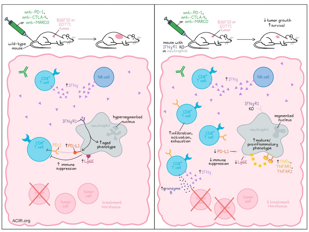
Investigating what happens to neutrophils unable to upregulate PD-L1 in tumors, Pei et al. found that upon treatment with immunotherapy, these cells showed increased infiltration and expressed higher levels of CD62L, Ly6E, CD11b, and CD101, maintaining their mature phenotype. They also expressed higher levels of TNFα, TNFAR1, and TNFAR2, suggestive of potential autocrine signaling and a more pro-inflammatory phenotype. With anti-CTLA-4 treatment, results were similar, though CD62L was downregulated. Looking at nuclear morphology, the researchers found that neutrophils unable to express PD-L1 maintained a segmented, mature phenotype following anti-PD-1, while those capable of upregulating PD-L1 developed a hypersegmented, aged nuclear morphology.
To determine whether interfering with IFNγ sensing by neutrophils might prevent the downstream PD-L1 upregulation and development of a more immunosuppressive phenotype, the researchers generated a mouse in which IFNγR1 was not expressed in neutrophils. Ex vivo analysis confirmed that these cells did not upregulate PD-L1 upon exposure to IFNγ, and in vivo analysis confirmed that immunotherapies showed more antitumor efficacy and prolonged survival in this model compared to in wild-type mice. In addition to the loss of IFNγ sensing and not upregulating PD-L1, neutrophils also showed reduced upregulation of markers associated with the more aged phenotype, and instead showed more pro-inflammatory potential and reduced mobility, which aligned with the increased accumulation and persistence of neutrophils in tumors. Some anti-PD-1-induced phenotypic alterations were also evident in neutrophils in the periphery.
Turning towards the potential human relevance of this research, Pei et al. analyzed scRNAseq data from human neutrophils cultured in different IFNs, and found that the results were similar to those observed in murine neutrophils. In scRNAseq data taken directly from patient NSCLC samples, neutrophils showed high Cd274 (PD-L1) expression, which was further increased in patients treated with anti-PD-1 combined with chemotherapy, and was higher in responders than non-responders, suggesting that increased PD-L1 likely resulted from a strong T cell-mediated IFNγ response. UMAP analysis revealed evidence that human neutrophils shifted towards an aged/immunosuppressive phenotype upon treatment. Neutrophils also showed enrichment for interferon response-related pathways and a trajectory towards an aged phenotype.
Overall, these results suggest that IFNγ induced in both murine and human tumors following certain immunotherapies is sensed by neutrophils, causing them to upregulate PD-L1 and polarize towards a more aged and immunosuppressive phenotype that limits antitumor responses. Targeting this mechanism could serve to alleviate this immunosuppression and improve responses to immunotherapy.
Write-up and image by Lauren Hitchings


